Ostium Secundum Atrial Septal Defect: A Case Report with Review of Literature
Saravanan Jagadeesan , Thirupathirao Vishnumukkala , Narendiran Krishnasamy , Shajan Koshy , Mohamad Aris Bin Mohd Moklas , Ahmad Yusuf Bin Yahaya · Int J Anat Res 2026;14(3):9607–9614
1 Department of Anatomy, School of Medicine, Lakeside Campus, Taylor’s University, Subang Jaya 47500, Selangor, Malaysia.
2 Anatomy Discipline, Human Biology Department, School of Medicine, IMU University, Bukit Jalil, Kuala Lumpur 57000, Malaysia.
3 Director, Clinical Skills Center, School of Medicine, Lakeside Campus, Taylor’s University, Subang Jaya 47500, Selangor, Malaysia.
4 Department of Human Anatomy, Faculty of Medicine and Health Sciences, Universiti Putra Malaysia, Serdang 43400, Selangor, Malaysia.
Corresponding Author: Dr. Ahmad Yusuf Bin Yahaya, Senior lecturer, Department of Anatomy, School of Medicine, Lakeside Campus, Taylor’s University, Subang Jaya 47500, Selangor, Malaysia. · Yusuf.Yahaya@taylors.edu.my
Abstract
Background
Atrial septal defect (ASD) is a congenital cardiac abnormality, representing roughly 10% of all congenital heart problems. The ostium secundum type is the truly familiar form, characterised by a deficit in the fossa ovalis. A significant proportion of instances, sometimes diagnosed in childhood, remain asymptomatic and undiscovered for the whole of a patient's life.
Methods
Case Presentation: An incidental discovery of a multi-fenestrated ostium secundum-type atrial septal defect was made during routine cadaveric dissection of a 60-year-old male cadaver. Five distinct fenestrations were observed within and around the fossa ovalis area of the interatrial septum, located in its superior, central, and inferior segments a configuration characteristic of the 'swiss-cheese' septal variety. The fossa ovalis boundary was discernible as a fibrous ring, however the floor and adjacent septum secundum tissue were significantly perforated. This individual has no recorded history of congenital heart disease.
Conclusion
This case highlights the clinical and anatomical importance of multi-fenestrated ostium secundum atrial septal defect, a morphological variety characterised by numerous distinct perforations instead of the anticipated singular central opening. The observed 'swiss-cheese' pattern has significant implications during surgical intervention, since it frequently prevents conventional single-device transcatheter closure and may require surgical patch repair. This case underscores the significance of cadaveric dissection in revealing intricate congenital abnormalities that may remain asymptomatic during a affected population life.
Background:
Congenital heart disorders (CHDs) constitute a diverse array of anatomical defects of the heart and major arteries resulting from defective embryogenesis which occurs during the initial eight weeks of intrauterine development [1]. Atrial septal defect (ASD) is one of the most common CHDs, with an incidence of roughly 1.6 per 1,000 live births, accounting for about 10% of all congenital cardiac malformations. [2]. ASD is characterised by abnormal communication between the two atria, allowing oxygenated blood to flow from the left atrium to the right atrium, resulting in right-sided volume overload and, ultimately, pulmonary hypertension and right heart failure [3].
ASD are categorised into four primary subtypes according to their anatomical position of the defect: (1) ostium secundum type, situated in the central area of the interatrial septum at the fossa ovalis; (2) ostium primum type, located in the inferior segment of the atrial septum contiguous to the atrioventricular valves; (3) sinus venosus type, sited near the entrance of the superior or inferior vena cava; and (4) coronary sinus type, the utmost uncommon variant. The ostium secundum ASD is the very familiar type, representing 75–80% of all ASDs [4,5].
A distinctive and clinically significant characteristic of ostium secundum ASD is its propensity to remain asymptomatic for decades, and in certain instances, throughout the patient’s life [6]. This is especially observed in the older demographic, where undetected ASDs may be discovered incidentally either during echocardiographic assessments for unrelated ailments, cardiac catheterisation, or, as in the current instance, after post-mortem cadaveric dissection [7].
The embryological origin of this defect has been thoroughly documented during cardiac development, the foramen ovale formed by the communication of septum primum and septum secundum fails to close entirely, or the septum secundum develops with excessive fenestration or insufficient tissue mass, resulting in a persistent interatrial communication. A big, centrally located defect corresponds to the ostium secundum phenotype [8].
We document an inadvertent finding of a substantial ostium secundum-type ASD in a sixty-year-old male cadaver during anatomical dissection. This represents valuable discovery that needs to be documented, considering the age of the cadaver and the lack of any previous clinical diagnosis. This paper offers an exhaustive examination of the literature related to the morphology, embryology, epidemiology, clinical manifestations, complications, and therapy of this illness.
CASE PRESENTATION
In the normal dissection session held in the Department of Anatomy, a male cadaver approximately 60 years old was examined as part of the standard instructional program. The cadaver was preserved utilising conventional embalming techniques with a 10% formalin solution. No notable medical history or cause of death was provided in the documents accompanying the cadaver.
Upon accessing the thoracic cavity and conducting a systematic dissection of the pericardium, the heart was in situ and subsequently excised according to established anatomical technique. The external inspection of the heart showed no apparent gross abnormalities, including the heart’s size and weight were normal.
Following a meticulous incision of the right atrium along the sulcus terminalis, the inside of the right atrium was revealed. The examination of the interatrial septum revealed numerous distinct perforations concentrated within fossa ovalis and next to the limbus fossa ovalis, indicative of a multi-fenestrated ostium secundum-type ASD (Figure 1). Five distinct fenestrations were identified: (1) a superior-left perforation in the upper-left quadrant of the fossa ovalis; (2) a superior-central fenestration situated just above the center of the fossa ovalis; (3) a smaller superior-right fenestration; (4) a mid-level left-margin perforation along the left border of the septum secundum; and (5) an inferior-left fenestration in the lower-left area of the fossa ovalis. The edges of each defect were smooth, hard, and fibrous, indicative of chronic organised perforations. No other structural anomalies were detected. The tricuspid and mitral valves appeared macroscopically normal.
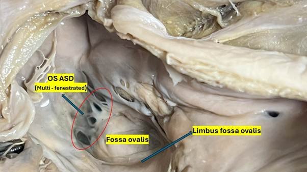
Fig. 1: Gross anatomical specimen of the right atrium illustrating a Mult fenestrated ostium secundum atrial septal defect (OS ASD). The interatrial septum is observed from the perspective of the right atrium. Numerous distinct fenestrations are evident within the fossa ovalis, indicative of a Mult fenestrated OS ASD, as highlighted in red. The next elevated muscle border limbus fossa ovalis (annulus ovalis) is well-preserved and complete.
The findings were consistent with a multi-fenestrated ostium secundum- ASD, a morphological variant characterised by excessive and spatially distributed resorption of the septum secundum, resulting in multiple discrete communications instead of a single large defect. This variation is very significant from both a morphological and clinical treatment standpoint.
DISCUSSION
Embryological basis of Ostium Secundum Atrial Septal Defect
Comprehending the embryology of the interatrial septum is essential for understanding the aetiology of ostium secundum ASD. Cardiac septation commences approximately in the fourth week of embryonic development [9]. The initial structure to develop is the septum primum, a crescent-shaped membrane barrier that extends downward from the ceiling of the primitive atrium near the endocardial cushions. As it develops, it forms a transient aperture known as the ostium primum at its lower edge [10].
Earlier to the closure of the ostium primum, programmed caspase-mediated cell death in the above segment of the septum primum generates a new aperture known as the ostium secundum, preserving interatrial connection vital for foetal circulation. Subsequently, the septum secundum, composed of more muscular and less flexible tissue, develops to the right of the septum primum and lowers, partially obscuring the ostium secundum. This produces a constricted opening referred to as the foramen ovale. [11].
In embryonic circulation, blood traverses from the right atrium to the left atrium through the foramen ovale, bypassing the inactive unborn lungs. Postnatally, with the commencement of pulmonary circulation and the increase in left atrial pressure, the septum primum is opposed to the septum secundum, thereby occluding the foramen ovale [12]. In roughly 75% of people, this functional closure is succeeded by anatomical fusion within the initial two years of life [13].
The ostium secundum ASD occurs when: (1) the ostium secundum is enlarged due to excessive apoptosis; (2) the septum secundum inadequately develops to cover the ostium secundum; or (3) there is abnormal resorption of the septum primum, leading to multiple fenestrations that merge into a significant central defect. The outcome is a sustained, haemodynamically relevant interatrial connection at the fossa ovalis [11].
Morphological Classification and the Multi-Fenestrated Variant
Ostium secundum ASD demonstrate significant morphological diversity. Classification can be founded on size (small <10 mm, moderate 10–20 mm, large >20 mm); shape (round, oval, irregular, or fenestrated); margin features (adequate rims against deficient rims in one or more sectors); and multiplicity (single defect versus numerous fenestrations) [14].
The multi-fenestrated ostium secundum ASD, as noted in this example, represents a unique and clinically significant morphological variant. This variety features several discrete perforations distributed throughout the fossa ovalis and the adjacent septum secundum tissue, rather than a singular big central connection [15]. The embryological foundation is thought to be due to spatially scattered excessive apoptosis in the septum primum and/or abnormal development of the overlaying septum secundum, leading to many residual holes that do not merge into a singular defect or achieve spontaneous closure [16].
In this cadaver, five fenestrations were observed, located in the superior, central, and inferior regions of the fossa ovalis. This distribution, encompassing all quadrants of the fossa ovalis, aligns with widespread rather than localised tissue resorption and closely mimics the ‘swiss-cheese’ septal variant documented in the literature [17].
The defect margins, known as rims, are clinically important for device closure planning. The rims consist of the aortic rim, the superior border (next to the superior vena cava), the inferior border (adjacent to the inferior vena cava), the posterior border, the atrioventricular edge (adjacent to the atrioventricular valves), and the coronary sinus rim. The multi-fenestrated form frequently lacks sufficient rim tissue between individual fenestrations, which renders transcatheter closure with a single device technically difficult or unfeasible necessitating open surgical patch repair [18]. This has obvious consequences for the clinical management of these patients and emphasises the necessity of precise morphological description using transoesophageal echocardiography or cardiac MRI before planning any intervention [19].
Haemodynamic Implications
The physiological effects of an ostium secundum ASD are mostly determined by the defect’s size and the accordance of the two ventricles. In cases of substantial left-to-right by-pass causes volume overload in the right atrium and right ventricle, resulting in continuing enlargement and hypertrophy of the right heart. The pulmonary vasculature experiences elevated flow, which over decades may result in structural alterations identified as pulmonary arterial hypertension (PAH) [20].
The shunt ratio (Qp:Qs), indicating the proportion of pulmonary to systemic blood flow, is a crucial factor in assessing severity. A ratio exceeding 1.5:1 is typically regarded as haemodynamically important. Untreated big atrial septal defects (ASDs) can progress to Eisenmenger syndrome, characterised by irreversible pulmonary hypertension and shunt reversal; however, this occurrence is less frequent in isolated ASDs than in ventricular septal defects [21].
Clinical Manifestation and Natural Progression in Adults
The clinical progression of ostium secundum ASD in adults is inconsistent. A significant number of patients remain asymptomatic until their third or fourth period of life, at which point symptoms such as exertional dyspnoea, tiredness, and palpitations occurs. Post-40 years of age, the occurrence of atrial fibrillation significantly increases in individuals with unrepaired ASD, due to atrial dilation and fibrosis caused by mechanical stretching [22].
Other notable consequences in elderly individuals with unrepaired ASD include: (1) paradoxical embolism via the defect, potentially resulting in stroke; (2) right heart failure; (3) supraventricular arrhythmias; (4) increasing pulmonary hypertension; and (5) infective endocarditis. A study by Campbell (1970) anticipated that 50% of individuals with large unrepaired ASDs would succumb before the age of 40; however, advancements in diagnostic techniques have since demonstrated that a considerable number of adults live into older age with moderate symptoms or remain fully asymptomatic [23].
The discovery of a 60-year-old individual with a significant, likely unrepaired ostium secundum atrial septal defect in this cadaver signifies that this could have been a clinically asymptomatic ASD or person suffered mild cardiovascular symptoms misattributed to other reasons.
Diagnostic Techniques
The diagnosis of ASD in clinical practice depends on a combination of clinical examination, electrocardiography, chest radiography, and echocardiography. Transthoracic echocardiography (TTE) functions as the principal diagnostic instrument, illustrating the defect, its dimensions, and the direction and magnitude of shunting by colour-flow Doppler. Transoesophageal echocardiography (TEE) offers enhanced resolution for rim evaluation, when planning for surgical closure [22].
Cardiac catheterisation, although predominantly replaced by echocardiography for diagnostic purposes, has its significance when evaluating pulmonary vascular resistance. Cardiac MRI is becoming a significant supplementary tool, providing accurate volumetric assessment of shunt flow and right ventricular performance. CT angiography is little utilised however can offer comprehensive anatomical insights [24].
Electrocardiogram findings with ostium secundum ASD typically exhibit right axis deviation, right bundle branch block (either incomplete or complete), and a longer PR interval in certain instances. Chest radiography may show cardiomegaly with right heart prevalence and increased pulmonary vascular patterns [25].
Management
The management method for ostium secundum ASD is based on the patient’s age, symptomatology, shunt ratio, pulmonary vascular resistance, and defect anatomy. The prevailing recommendations from the American College of Cardiology/American Heart Association (ACC/AHA) and the European Society of Cardiology (ESC) advocate for the closure of haemodynamically significant atrial septal defects (ASDs) (Qp:Qs ≥1.5:1) in all symptomatic individuals and in asymptomatic individuals who exhibit signs of right heart volume overload [26].
Two primary methodologies are accessible: transcatheter device occlusion and surgical intervention. Transcatheter closure using Amplatzer Septal Occluder or analogous devices has emerged as the preferred method for anatomically appropriate ostium secundum ASD, providing reduced procedural risk, lower duration of hospitalisation, and comparable efficacy to surgical intervention. The gold standard for big lesions with inadequate rims, intricate morphology, or related anomalies is surgical closure with direct suture or patch repair [27].
Significantly, in elderly patients (>60 years), ASD closure has demonstrated enhancements in functional capability and excellence of life, a reduced frequency of new-onset atrial fibrillation, and a potential increase in lifespan, provided there is absence of irreversible pulmonary arterial hypertension. These observations underscore the significance of not disregarding incidental ASD findings in the elderly as trivial [26].
Importance of Cadaveric Observations in Anatomical Education
The identification of substantial circulatory irregularities during cadaveric dissection possesses considerable educational and scientific significance. These findings offer medical students a concrete, three-dimensional understanding of congenital defects that no textbook or digital medium can entirely reproduce. Furthermore, case studies detailing cadaveric anomalies enhance the anatomical and pathological literature by identifying morphological variances and their potential clinical implications [28].
Prior case reports have detailed different cardiac abnormalities unexpectedly discovered in cadavers, such as bicuspid aortic valves, persisting left superior vena cava, aberrant coronary artery origins, and ventricular septal defects [29, 30]. The documentation of ostium secundum ASD in a 60-year-old male cadaver significantly contributes to the existing knowledge, since it illustrates that substantial abnormalities can persist undiscovered in a functionally normal man for more than sixty years.
CONCLUSION
We report an uncommon incidental discovery of a multi-fenestrated ostium secundum-type ASD in a 60-year-old male cadaver identified during standard anatomical dissection. Five distinct openings were observed within and surrounding the fossa ovalis, located across the superior, central, and inferior regions of the interatrial septum having morphological characteristics of a multi-fenestrated pattern of ASD which has been a clinically unrecognised congenital cardiac abnormality.
This case underscores several critical educational points: firstly, multi-fenestrated ostium secundum ASD can be asymptomatic and undiagnosed into advanced age; secondly, the ‘swiss-cheese’ morphological configuration observed here has specific ramifications for transcatheter closure, often requiring surgical patch repair instead of single-device deployment; thirdly, the anatomical dissection room serves as an invaluable venue for the identification and documentation of intricate congenital anomalies that enhance both undergraduate medical education and the published anatomical corpus.
We advocate for the systematic documentation and reporting of all accidental findings in cadaveric dissection to enhance the understanding of undetected congenital heart disease among the general population. Future research involving post-mortem cardiac evaluations with systematic documentation may assist in determining the actual prevalence of undiagnosed ASD in the older population.
ACKNOWLEDGEMENTS
The authors like to express gratitude to the cadaver donors and their families, whose altruistic donation to medical science facilitates anatomical instruction and research of this kind.
Conflicts Of Interest: The authors assert the absence of any conflicts of interest with the publishing of this manuscript.
Funding: This research did not receive any specific support from agencies.
Author Contribution
Saravanan Jagadeesan: Conceptualization of the cadaveric case report, performed cadaveric dissection, and drafted the manuscript. Thirupathirao Vishnumukkala: Interpreted anatomical findings, literature findings, and critically revised the manuscript. Narendiran Krishnasamy: Participated in specimen examination, literature review, and manuscript editing. Shajan Koshy: Supervised the anatomical study and dissection procedures and reviewed the manuscript. Mohamad Aris Bin Mohd Moklas: Interpretation of anatomical findings, literature review and reviewed the manuscript. Ahmad Yusuf Bin Yahaya: Performed dissection, provided overall supervision, contributed to study design and critical manuscript revision, and approved the final manuscript for publication. All authors contributed substantially to the preparation of this manuscript and approved the final version for publication.
Cite this article: Saravanan Jagadeesan, Thirupathirao Vishnumukkala, Narendiran Krishnasamy, Shajan Koshy, Mohamad Aris Bin Mohd Moklas, Ahmad Yusuf Bin Yahaya. Ostium Secundum Atrial Septal Defect: A Case Report with Review of Literature. Int J Anat Res. 2026;14(3):9607–9614. DOI: 10.16965/ijar.2026.161
References
- Meng, X., Song, M., Zhang, K., Lu, W., Li, Y., Zhang, C., & Zhang, Y. Congenital heart disease: types, pathophysiology, diagnosis, and treatment options. MedComm, 2024;5(7):e631. https://doi.org/10.1002/mco2.631 PMid:38974713 PMCid:PMC11224996
- Xu, J., Li, Q., Deng, L., Xiong, J., Cheng, Z., & Ye, C. Global, regional, and national epidemiology of congenital heart disease in children from 1990 to 2021. Frontiers in cardiovascular medicine, 2025;12:1522644. https://doi.org/10.3389/fcvm.2025.1522644 PMid:40454242 PMCid:PMC12122482
- Bartoszewska, E., Chrapkowska, A., Zielińska, O., Mordalska, M., Lizon, J., Zalewska, Z., & Wasicionek, M. Atrial Septal Defects: From Embryology to Pediatric Pulmonary Hypertension. Journal of clinical medicine, 2025;14(21):7698. https://doi.org/10.3390/jcm14217698 PMid:41227094 PMCid:PMC12609445
- Hoffman JI, Kaplan S. The incidence of congenital heart disease. J Am Coll Cardiol. 2002;39(12):1890-1900. doi:10.1016/s0735-1097(02)01886-7 https://doi.org/10.1016/S0735-1097(02)01886-7 PMid:12084585
- Webb G, Gatzoulis MA. Atrial septal defects in the adult: recent progress and overview. Circulation. 2006;114(15):1645-1653. https://doi.org/10.1161/CIRCULATIONAHA.105.592055 PMid:17030704
- Ejim, E. C., Anisiuba, B. C., Ike, S. O., & Essien, I. O. Atrial septal defects presenting initially in adulthood: patterns of clinical presentation in enugu, South-East Nigeria. Journal of tropical medicine, 2011;251913. https://doi.org/10.1155/2011/251913 PMid:21760805 PMCid:PMC3134101
- Angelini, A., di Gioia, C., Doran, H., Fedrigo, M., Henriques de Gouveia, R., Ho, S. Y., Leone, O., Sheppard, M. N., Thiene, G., Dimopoulos, K., Mulder, B., Padalino, M., van der Wal, A. C., & Association for European Cardiovascular Pathology (AECVP). Autopsy in adults with congenital heart disease (ACHD). Virchows Archiv : an international journal of pathology, 2020;476(6):797-820. https://doi.org/10.1007/s00428-020-02779-8 PMid:32266476 PMCid:PMC7272495
- Briggs, L. E., Kakarla, J., & Wessels, A. The pathogenesis of atrial and atrioventricular septal defects with special emphasis on the role of the dorsal mesenchymal protrusion. Differentiation; research in biological diversity, 2012;84(1):117-130. https://doi.org/10.1016/j.diff.2012.05.006 PMid:22709652 PMCid:PMC3389176
- Oliveira, R. C., Martins, P., & Martins, M. F. The Congenital Malformation of the Interatrial Septum-A Review of Its Development and Embryology with Clinical Implications. Journal of developmental biology, 2025;13(3):28. https://doi.org/10.3390/jdb13030028 PMid:40843896 PMCid:PMC12372156
- Naqvi N, McCarthy KP, Ho SY. Anatomy of the atrial septum and interatrial communications. J Thorac Dis 2018;10(Suppl 24):S2837-S2847. https://doi.org/10.21037/jtd.2018.02.18 PMid:30305943 PMCid:PMC6174145
- Nayak S. B. Functional and clinical importance of a large sized ostium secundum defect in a middle aged female cadaver: a case report. Anatomy & cell biology, 2017;50(2):152-154. https://doi.org/10.5115/acb.2017.50.2.152 PMid:28713619 PMCid:PMC5509899
- Zhang, D., & Lindsey, S. E. Recasting Current Knowledge of Human Fetal Circulation: The Importance of Computational Models. Journal of cardiovascular development and disease, 2023;10(6):240. https://doi.org/10.3390/jcdd10060240 PMid:37367405 PMCid:PMC10299027
- Kutty S, Sengupta PP, Khandheria BK. Patent foramen ovale: the known and the to be known. J Am Coll Cardiol. 2012;59(19):1665-1671. https://doi.org/10.1016/j.jacc.2011.09.085 PMid:22554596
- Warnes CA, Williams RG, Bashore TM, et al. ACC/AHA 2008 guidelines for the management of adults with congenital heart disease: a report of the American College of Cardiology/American Heart Association Task Force on Practice Guidelines (Writing Committee to Develop Guidelines on the Management of Adults With Congenital Heart Disease). Developed in Collaboration With the American Society of Echocardiography, Heart Rhythm Society, International Society for Adult Congenital Heart Disease, Society for Cardiovascular Angiography and Interventions, and Society of Thoracic Surgeons. J Am Coll Cardiol. 2008;52(23):e143-e263. https://doi.org/10.1161/CIRCULATIONAHA.108.190690 PMCid:PMC5408160
- McCarthy, K., Ho, S., & Anderson, R. Defining the morphologic phenotypes of atrial septal defects and interatrial communications. Images in paediatric cardiology, 2003;5(2):1-24. Al Hennawi, H., Bandi, A., Abulshamat, A., Srivastava, S., & Qintar, M. (2024). Transcatheter Closure of Complex Multifenestrated Atrial Septal Defect Using Three Septal Occluders: Is Three Too Many?. Structural heart: the journal of the Heart Team, 2024;9(3):100355. https://doi.org/10.1016/j.shj.2024.100355 PMid:40212336 PMCid:PMC11979385
- Al Hennawi, H., Bandi, A., Abulshamat, A., Srivastava, S., & Qintar, M. (2024). Transcatheter Closure of Complex Multifenestrated Atrial Septal Defect Using Three Septal Occluders: Is Three Too Many?. Structural heart: the journal of the Heart Team, 2024;9(3):100355. https://doi.org/10.1016/j.shj.2024.100355 PMid:40212336 PMCid:PMC11979385
- Kao, A. S., & Cardozo, S. Three-Dimensional Cardiac Imaging: A Necessity to the Diagnosis and Treatment of Swiss-Cheese Atrial Septal Defect. Cureus, 2023;15(6): e40341. https://doi.org/10.7759/cureus.40341
- Masura J, Gavora P, Formanek A, Hijazi ZM. Transcatheter closure of secundum atrial septal defects using the new self-centering amplatzer septal occluder: initial human experience. Cathet Cardiovasc Diagn. 1997;42(4):388-393. https://doi.org/10.1002/(SICI)1097-0304(199712)42:4<388::AID-CCD7>3.0.CO;2-7
- Baumgartner H, De Backer J, Babu-Narayan SV, et al. 2020 ESC Guidelines for the management of adult congenital heart disease. Eur Heart J. 2021;42(6):563-645.
- Van De Bruaene A, Delcroix M, Pasquet A, et al. The importance of pulmonary artery pressures on late atrial arrhythmia in transcatheter and surgically closed ASD type secundum. Int J Cardiol. 2011;152(2):192-195. https://doi.org/10.1016/j.ijcard.2010.07.014 PMid:20674051
- Torres A. J. Hemodynamic assessment of atrial septal defects. Journal of thoracic disease, 2018;10(Suppl 24):S2882-S2889 https://doi.org/10.21037/jtd.2018.02.17 PMid:30305948 PMCid:PMC6174148
- Martin, S. S., Shapiro, E. P., & Mukherjee, M. Atrial septal defects - clinical manifestations, echo assessment, and intervention. Clinical Medicine Insights. Cardiology, 2015;8(Suppl 1):93-98. https://doi.org/10.4137/CMC.S15715 PMid:25861226 PMCid:PMC4373719
- Dehghani, H., & Boyle, A. J. Percutaneous device closure of secundum atrial septal defect in older adults. American journal of cardiovascular disease, 2012;2(2): 133-142. Katbamna B, Wu L, Rodriguez M, et al. The uses of right heart catheterization in cardio-pulmonary disease: State-of-the-art. Am Heart J Plus. 2024;49:100488. Published 2024 Dec 6. https://doi.org/10.1016/j.ahjo.2024.100488 PMid:39760109 PMCid:PMC11699050
- Katbamna B, Wu L, Rodriguez M, et al. The uses of right heart catheterization in cardio-pulmonary disease: State-of-the-art. Am Heart J Plus. 2024;49:100488. Published 2024 Dec 6. https://doi.org/10.1016/j.ahjo.2024.100488 PMid:39760109 PMCid:PMC11699050
- Kang, J. H., Wu, H. Y., & Long, W. J. Effects of atrial septal defects on the cardiac conduction system. World journal of clinical cases, 20024;12(35):6770-6774. https://doi.org/10.12998/wjcc.v12.i35.6770 PMid:39687649 PMCid:PMC11525910
- Stout KK, Daniels CJ, Aboulhosn JA, et al. 2018 AHA/ACC Guideline for the Management of Adults With Congenital Heart Disease: Executive Summary: A Report of the American College of Cardiology/American Heart Association Task Force on Clinical Practice Guidelines. J Am Coll Cardiol. 2019;73(12):1494-1563.
- Kashyap, T., Sanusi, M., Momin, E. S., Khan, A. A., Mannan, V., Pervaiz, M. A., Akram, A., & Elshaikh, A. O. Transcatheter Occluder Devices for the Closure of Atrial Septal Defect in Children: How Safe and Effective Are They? A Systematic Review. Cureus, 2022;14(5):e25402. https://doi.org/10.7759/cureus.25402
- Ghazanfar, H., Rashid, S., Hussain, A., Ghazanfar, M., Ghazanfar, A., & Javaid, A. (2018). Cadaveric Dissection a Thing of the Past? The Insight of Consultants, Fellows, and Residents. Cureus, 2018;10(4):e2418. https://doi.org/10.7759/cureus.2418
- Dave, V., Sesham, K., Mehra, S., Roy, T. S., & Ahuja, M. S. Persistent left superior vena cava: An anatomical variation. Medical journal, Armed Forces India, 2022;78(Suppl 1):S277-S281. https://doi.org/10.1016/j.mjafi.2020.01.009 PMid:36147413 PMCid:PMC9485778
- Vallabhajosyula, S., Fuchs, M., Yang, L. T., Medina Inojosa, J., Tajouri, T. H., Enriquez-Sarano, M., Phillips, S. D., Gulati, R., Klarich, K. W., & Michelena, H. Anomalous coronary artery origin from the opposite sinus in patients with bicuspid aortic valve: comparison with tricuspid aortic valve. Open Heart, 2021;8(1):e001567. https://doi.org/10.1136/openhrt-2020-001567 PMCid:PMC8217920


