
Impact Of Caffeine on the Neurodegenerative Changes of the Frontal Cortex in Type 2 Diabetic Rats
Manal A. Othman, Ayesha Fatima , Bello Sirajo , Mohammed Khan , Raouf A. Fadel · Int J Anat Res 2026;14(3):9599–9606
1 Department of Anatomy, College of Medicine and Health Sciences, Arabian Gulf University, Manama, Bahrain.
2 Department of Histology and Cell Biology, Faculty of Medicine, Assiut University, Assiut, Egypt.
3 Department of Human Anatomy and Embryology, Faculty of Medicine, Suez Canal University, Ismailia, Egypt.
Corresponding Author: Manal A. Othman, Department of Anatomy, College of Medicine and Health Sciences, Arabian Gulf University, Manama, Bahrain. Phone +97317239507 P.O Box: 26671, Manama, Kingdom of Bahrain. · manalamo@agu.edu.bh
Abstract
Background
Type 2 diabetes (T2D) is associated with injury to many organs in our body. The central nervous system can be affected in diabetes progression, which may result in dementia and decline in cognitive function. Myelin is very important in neuronal protection and axonal transport and might be affected with neurodegenerative diseases as diabetes. Caffeine, a commonly used psychoactive beverage, is known to improve the impairment in memory and cognitive function. This work was aiming at investigating the protective role that caffeine can provide for T2D rat neurodegeneration.
Methods
Thirty-six male Wistar rats, were grouped into 4 groups (9 each); the control group (group 1), diabetic group (group 2), caffeine + diabetic group (group 3), and caffeine-only group (group 4). Induction of T2D was done by feeding the rats with high calorie diet and giving them a single injection of streptozotocin intraperitoneally at a lower dose. Caffeine was administered by oral route for 5 weeks. The rats were sacrificed; brains were removed and put in 10% formalin fixative and processed to evaluate their general morphology. Immunohistochemistry was performed using neuronal, myelin and apoptotic markers.
Results
Histological examination revealed neurodegeneration of frontal cortex of rats with T2D. Immunohistochemistry revealed downregulation in neuronal and myelin immunostaining and upregulation of apoptosis in rats with T2D.
Conclusion
There was enhancement of the structure and immunohistochemistry of frontal cortex of diabetic rats, which receive caffeine. This neuroprotective effect of caffeine in T2D rats suggests a therapeutic potential for caffeine in T2D.
BACKGROUND
Type 2 diabetes (T2D) is considered a lifelong metabolic disease affecting many organ systems, including the central nervous system [1]. It is characterized by hyperglycemia due to insulin resistance in peripheral tissues and a persistent decline in the function of β cells of the pancreas. The use of diets with high calories, and streptozotocin (STZ) at lower doses, can generally be accepted to produce a rodent model of T2D [2,3]. Persistent hyperglycemia can gradually disrupt normal brain structure and function, which contributes to cognitive decline, cerebral atrophy, infarction, and neurodegeneration [4,5].
The frontal cortex is a major region of the brain responsible for thinking, decision-making, and overall cognitive function. Evidence from previous studies shows that chronic T2D, characterized by hyperglycemia, insulin resistance, and microvascular injury, gradually causes changes in the structure and function of this vital region of the brain [6]. Neuroimaging studies in long-standing diabetics reveal reduced frontal lobe grey matter volume and thinning of the frontal cortex, indicating increased neurodegeneration in this region [7,8]. In a mouse model of T2D, researchers found not only reductions in the overall size of the brain and cortex, but also changes in the cellular composition of the cortex, such as a decrease in the number of mature neurons, impaired myelination, and disruptions to vascular and glial cell structure. These findings suggest that diabetes can impair the neurovascular unit, degrade neural and glial integrity, and compromise brain microstructure, thereby contributing to cognitive decline [9]. We previously reported that rats administered a high-calorie diet showed defective structure in the hippocampus, which was mediated by gliosis and inflammation [10].
Caffeine is a naturally occurring psychostimulant that is globally consumed in the form of drinks like coffee and tea. It produces increased alertness, reduced fatigue, and elevated mood by blocking adenosine receptors [11]. Recent research shows that caffeine has neuroprotective, antioxidant, and anti-inflammatory effects, thus decreasing the risk of degenerative diseases as Parkinson’s and Alzheimer disease. This is likely due to mechanisms including reduction of oxidative stress, modulation of adenosine receptor signaling, and suppression of neuroinflammation [12].
Diabetes can progress over time and might lead to severe morbidities and mortalities. The pathophysiology of the brain damage in diabetic encephalopathy is not fully understood. The current study aimed at investigating the possibility of caffeine to improve neurodegeneration in the frontal cortex of T2D rats through the investigation of neuronal affection, myelination, and apoptosis.
Materials and Methods
Animals: Adult male Wistar rats, weighing 270-300 g, were chosen for the study; they were placed in the animal house facility, at the college of Medicine and health sciences, Arabian Gulf University, Bahrain. They were placed at standard light/dark cycle at room temperature, and were given free access to tap water. The study was performed according to the Guide for the Care and Use of Laboratory animals of the National Institutes of Health (NIH). The experiments of this study were performed after approval of the Committee of Animal Care and Use at the Arabian Gulf University, Bahrain: ethical approval (# E006-I-04/17).
Induction of T2D and animal groups
The induction of T2D was previously established in our lab [10]. Briefly, 36 rats were divided into 4 groups (9 rats each). The rats of control group (1) were given diet with low calories (LCD) (SF 18-050, Specialty Feeds, Australia) and injected intraperitoneally (IP) with citrate buffer and given oral saline. The diabetic rats (group 2) were given a diet with high calories (HCD) (SF-04-001, Specialty Feeds, Australia). Four weeks later, rats receive streptozotocin (STZ) intraperitoneally (SigmaTM, St. Louis, MO, USA). STZ was dissolved in citrate buffer and given as a single dose of 35 mg/kg body weight (BW).
Three days after injection with STZ, blood samples were obtained from rat tails after fasting overnight, for blood glucose measurement. Animals that measured levels of blood glucose over 250 mg/dL were considered diabetic. Diabetic rats that receive caffeine (group 3): were administered caffeine at a dose of 100 mg/kg BW (Sigma TM, Saint Louis, MO, USA). Caffeine was given by oral route through gavage, after dissolving in saline, every day for five-week period. The caffeine-only group (group 4): were the rats that had LCD and 100 mg/kg BW of caffeine orally, every day for 5 weeks. After 8 weeks of the experiments, animals were sacrificed with inhalation of CO2 and their brains were dissected out and processed for histological and immunohistochemical evaluations.
Blood glucose measurements
The detection of blood glucose, from rats of all groups, was performed at the start of the study, 2 weeks after feeding with HCD and 72 hours after STZ injections, to warrant diabetic status. Furthermore, blood glucose measurement was performed every week after caffeine administration. Twelve hours after fasting, a drop of blood was withdrawn from rat tails (distal ends) and immediately measured using (Accu-Check Active, Roche Diagnostics, Mannheim, Germany).
Histological Evaluation
After sacrificing the animals, the brains were dissected out from all of them, and washed with saline. The samples were placed in the fixative (10% neutral buffered formalin). Then the samples including the frontal cortex, were trimmed, and processed to obtain blocks of paraffin. Five µm coronal sections were obtained by a rotating microtome. For evaluation of general histology, hematoxylin and eosin (Hx&E) staining was performed. The photo capture was done using the objective lens, 20X of a light microscope (Axioscope A1, Carl Zeiss Microscopy, Germany) which was connected with a digital camera. Histological processing and staining were performed following [13].
Immunohistochemical study
Five µm paraffin sections, were put on slides (charged) and immunohistochemistry was performed by an Avidin-Biotin detection kit (Vectastain Elite ABC Universal Kit, Vector Laboratories, UK) according to the instructions of the manufacturer. Incubation of the sections was done using the following primary antibodies, separately: neuronal marker, anti NeuN, a rabbit monoclonal antibody (ab177487, Abcam, UK), myelin marker, anti-myelin basic protein (MBP) a rabbit monoclonal antibody (ab218011, Abcam, UK), and an apoptotic marker, anti-Bax, a rabbit monoclonal antibody (ab32503, Abcam, UK), overnight in the refrigerator (4ºC).
Goat serum was used as non-immune negative control and 3,3 diaminobenzidine tetrahydrochloride to help visualizing the reaction. The sections were then put in Harris hematoxylin for counterstaining. Examination and photo capture of the slides were done by the light microscope. Quantitative findings of photomicrographs of immunohistochemistry markers, were analyzed by the software of image J® (Wayne Rasband NIH, Bethesda, MD, USA) on 6 overlapping fields of 6 slides for each animal.
Statistical analysis
Analysis of data was done by the use of the Statistical Package for Social Sciences® (SPSS) software version 27.0 for Windows (IBM Corp., Armonk, NY, USA). Expression of variables was the mean ± standard deviation (SD). Analysis of data was done using one way analysis of variance (ANOVA) and Tukey post hoc test. P value of less than 0.05 is considered statistically significant.
Results
Blood glucose level
Blood glucose values of all rats at the beginning of the study, were normal (85± 2.2 mg/dL). After HCD and 3 days of injection of STZ, there was a significant increase in the levels of blood glucose, for the injected rats which accomplished diabetic state and they remained with high levels through the whole experiment (303 ±15mg/dL, P< 0.0001). Caffeine administration decreased the levels of blood glucose in comparison to rats with diabetes (109 ± 2.7, P< 0.0001). Blood glucose levels of caffeine only treated animals had no significantly statistical difference in comparison to control rats (86 ±1.8, P=1.00) (Figure 1).
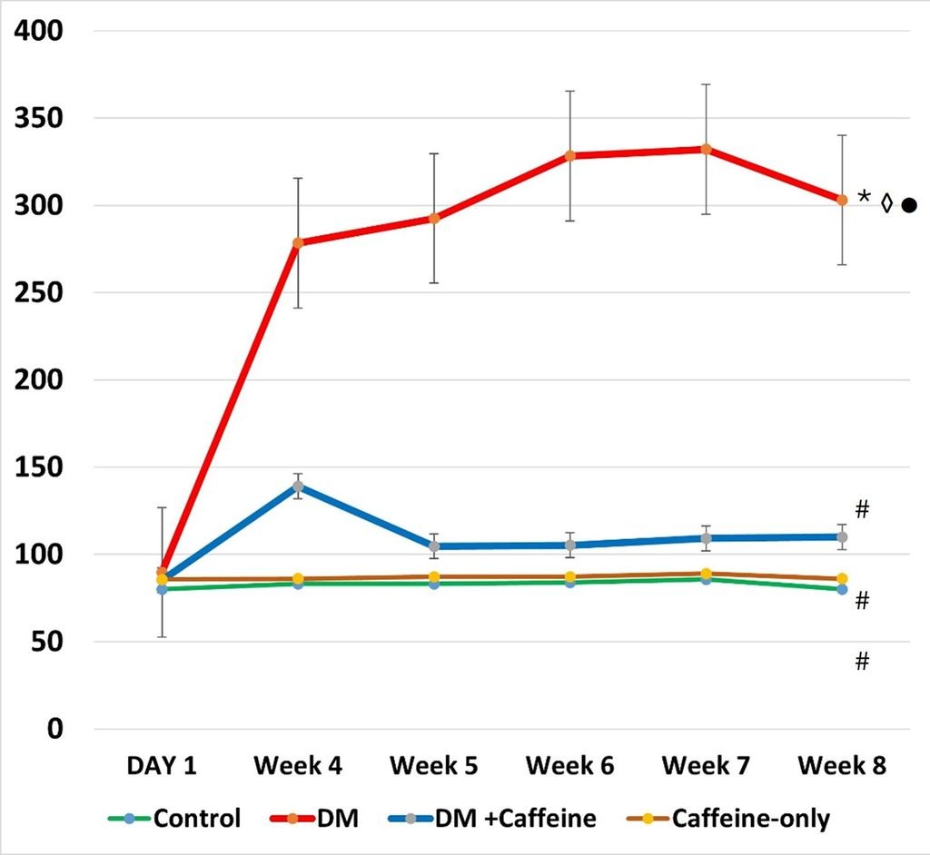
Fig. 1: Comparison of blood glucose levels among the different studied groups. Each value represents the mean of blood glucose levels at day 1, week 4, week 5, week 6, week 7 and week 8. The error bars present standard errors. # vs DM, vs control, vs DM + Caffeine, vs Caffeine-only, P < 0.0001.
Hx&E general morphology
Caffeine improved the neuronal distortion that occurred in diabetic animals
Evaluation of histological samples of the frontal cortex tissues in all experimental groups, was done by staining with Hx&E stain. The control group had intact neurons, which were well organized and having pale central nucleus and cytoplasm. In the frontal cortex of diabetic rats, there was numerous dark cells with pericellular spaces, vacuolization, in addition to pyknotic nuclei. In the caffeine treated diabetic rats, there was preservation of the neuronal structure with healthy appearing neurons and fewer darkly stained and vacuolated cells compared to diabetic group. Caffeine-only treated group showed normal cellular components and no neuronal disruption (Fig. 2).
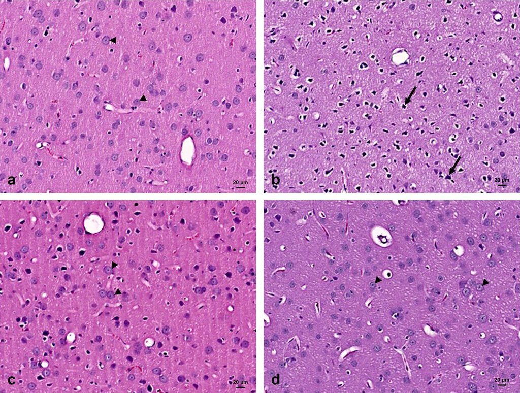
Fig. 2: Photomicrographs of the frontal cortex stained with Hx&E. A: Control section reveals the neurons with their pale cytoplasm and vesicular nucleus. B: The diabetic group shows that most of cells have dark cytoplasm and nucleus with pericellular halo. C: The caffeine treated diabetic group are showing many cells with pale cytoplasm and vesicular nuclei. There are few dark cells with dark nuclei. D: The caffeine group are showing mainly pale cells as well. X 200
Immunohistochemistry
Caffeine prevented diabetes‑induced neuronal loss
The neuronal cells in the frontal cortex were detected by NeuN immunohistochemistry of the frontal cortex. Diabetic rats showed decreased expression in comparison to the control group (p<0.001). Diabetic rats that received caffeine, showed marked increase in NeuN immunoreactivity (p˂0.001). Caffeine-only group showed levels like the control group (Fig. 3 & table 1).
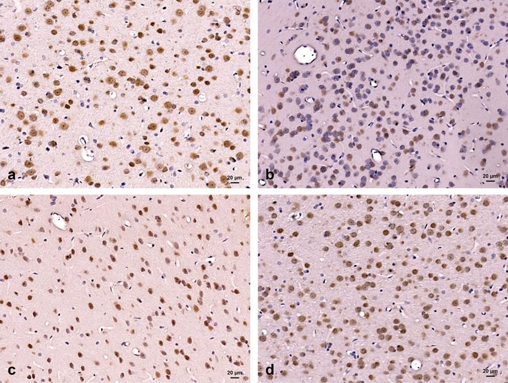
Fig. 3: Photomicrographs of the frontal cortex immunostained with anti-NeuN antibody (Neuronal marker). A: Control group is showing the immunoreactivity of neurons. B: Diabetic group displays a marked decrease in immunoreaction. C: Caffeine-treated diabetic group reveals a marked increase in immunoreactivity. D: Caffeine only treated animals show the positive staining of neurons. X 200.
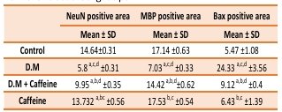
Table 1: Percent area of immunoreactivity in the different studied groups.
Standard deviation (SD), one way analysis of variance (ANOVA) was used followed by Tukey for multiple comparisons a: significant compared to control group, b significant compared to DM group, c significant compared to DM treated with Caffein group, d significant compared to caffein only group.
Caffeine prevented diabetes‑induced myelin downregulation
The myelin in neuronal cells of the frontal cortex was detected by MBP immunohistochemistry. Diabetic rats showed decreased expression compared to the control group (p<0.001). On the other hand, caffeine treated diabetic rats, revealed marked increase in MBP immunoreactivity (p˂0.001). Caffeine-only group showed values similar to the control group (Fig. 4 & table 1).
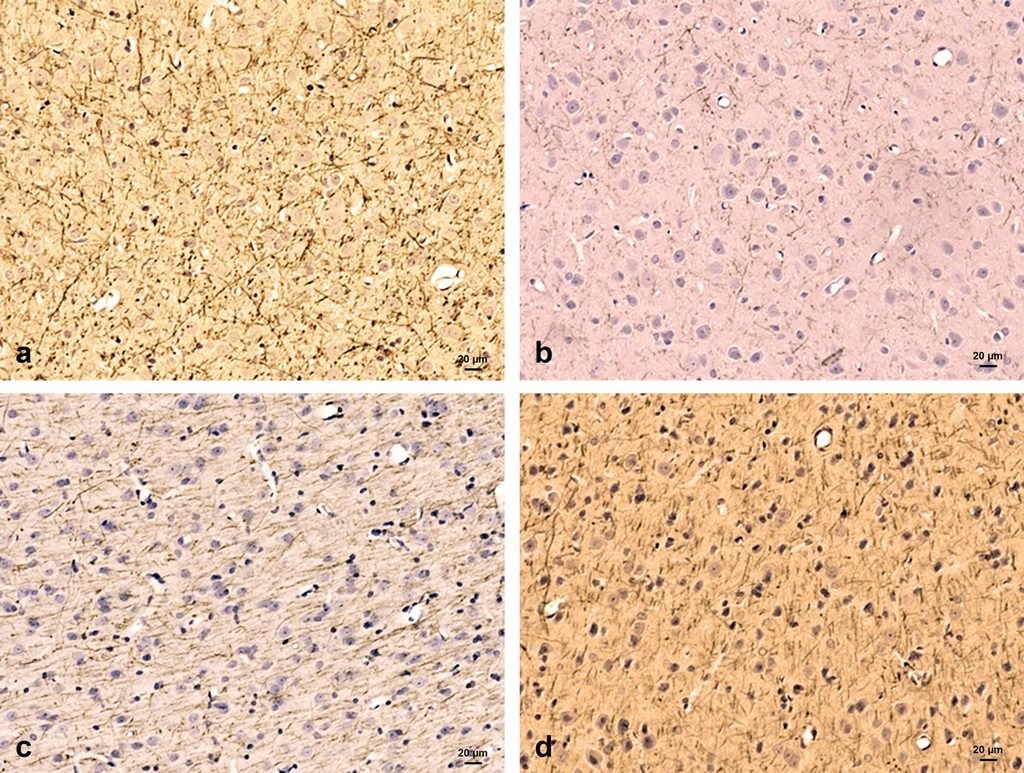
Fig. 4: Photomicrographs of the frontal cortex immunostained with anti-MBP antibody (Myelin marker). A: Control group shows the immunoreactivity of myelinated cells. B: Diabetic group shows a marked decrease in immunoreaction. C: Caffeine-treated diabetic group reveals a marked increase in immunoreactivity. D: Caffeine only treated animals show the positive staining of myelin. X 200
Caffeine decreased diabetes‑induced apoptosis
The apoptosis reaction was detected by Bax immunohistochemistry of the frontal cortex. Diabetic animals revealed marked expression in comparison to the control group (p<0.001). Caffeine-treated diabetic group displayed a significant decline in the immunostaining of Bax (p˂0.001). The group with caffeine only showed immunoreaction similar to control group (Fig. 5 & table 1).
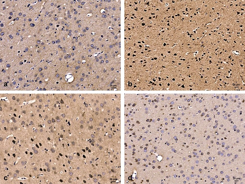
Fig. 5: Photomicrographs of the frontal cortex immunostained with anti-Bax antibody (Apoptosis marker). A: Control group shows no immunoreactivity of cells. B: Diabetic group shows a marked increase in immunoreaction. C: Caffeine-treated diabetic group reveals a decrease in immunoreactivity. D: Caffeine only treated animals show no staining similar to control. X 200
Discussion
This study examined the role of caffeine in protecting T2D-induced effects on the rat frontal cortex, in which T2D was induced by high calorie diet followed by STZ injection as previously established in our lab [10]. Caffeine administration decreased blood glucose levels and improved neurodegeneration. There are other studies that used similar models with different regimens [14,15]. After the administration of caffeine, the deleterious effects as hyperglycemia and neurodegeneration improved significantly. The frontal cortex was chosen to evaluate the findings of diabetes because the frontal cortex is a crucial region of the brain, which can be affected by complications of diabetes and its affection might result in cognitive and memory impairment [16,17].
In this study, histological evaluation of the frontal cortex of rats in group 2 (T2D group) showed many dark neurons with dark nuclei and vacuolated cytoplasm. Similar results were detected in the hippocampal tissues in type 1 diabetes, in our previous study [18] and by others (reviewed in [19]). Additionally, caffeine was found to decrease apoptosis in frontal cortical cells of T2D rats. This might denote that caffeine can ameliorate apoptosis in the frontal cortex in cases of diabetes as previously reported in protecting bladder neurological function in diabetic rats [20]. Recently, it was reported that caffeine decreased the retinal cells apoptosis induced by oxidative stress and this is mainly due to its antioxidative effect [21]. Apoptosis is important in neurodegenerative disease progress. Central nervous system affection in diabetes, might result in degenerative changes in the brain and neurovascular areas [22,23].
Neuronal loss was also demonstrated by NeuN immunohistochemistry, in this study, where the expression was less in T2D rats. A similar study reported neuronal loss and other neurodegenerative changes in monkeys with spontaneous T2D [24]. Furthermore, the neuronal loss in T2D frontal cortex, can be associated with insulin resistance and hyperglycemia, which may be related to the pathophysiology of cognitive dysfunctions [4,25].
Caffeine is known to be an antagonist to adenosine 1 receptor (A1R) and A2R. The effect of caffeine in preventing impairment of memory in T2D is mainly mediated by A2R receptor not A1R as the targeted inhibition of A2R leads to activation of caffeine’s effects (reviewed in [26]). A2Rs are concentrated in the frontal cortex and other brain areas, in which they modulate synaptic plasticity [27]. Moreover, antagonizing A2R receptor may result in great neuroprotective roles in a wide variety of CNS disorders [28].
This study demonstrated a decrease in myelin in the frontal cortex of diabetic rats, which is detected by MBP expression and a marked improvement after caffeine administration. This can align nicely with our previous finding of a similar result in the peripheral nerve of T2D, which was detected by MBP expression and electron microscopy [29]. Another study demonstrated a protective effect of caffeine and myelin enhancement in a hypoxic ischemic rat model [30]. Downregulation of the levels of MBP was reported in neurodegenerative conditions, like Parkinson’s and Alzheimer’s disease [31,32].
Since motor and cognitive function, which is a main function of the frontal cortex, is closely related to myelin formation, disturbance in the integrity and expression of myelin as detected in AD [33]. On the other hand, oligodendrocyte reaction to glucose toxicity remains largely unexplored. This study is contributing an insight into the association of myelin disruption to T2D, resulting from glucotoxicity in oligodendrocytes and the brain of this T2D rat model.
The results of the current study display myelin disruption in rats with T2D, in comparison to those treated with caffeine, which supports remyelination. Type 2 Diabetes is associated with hyperglycemia, and this may result in increased levels of glucose levels in neurons, impairing glucose metabolism intracellularly and can result in neuronal injury [34]. Previously, it was reported that increase glucose levels may lead to oxidative stress and neuroinflammation, which might result in apoptosis [35]. This is evidenced by Bax upregulation in this study and, elevated inflammatory cytokine expression like IL-6 and TNF-α, as previously reported [10].
Conclusion
Our study detected improvement of neurodegenerative changes of T2D rats by the consumption of caffeine. The findings in this study can provide an insight into the mechanisms of neurodegeneration of the frontal cortex in T2D, which is mediated by demyelination, neuronal loss and apoptosis. This protective effect of caffeine can support its use as a therapeutic modality in T2D. Future research will further investigate this therapeutic potential and unravel the complex pathophysiology of T2D, including behavioral testing and oxidative stress.
Abbreviations
T2D: type 2 diabetes, STZ: streptozotocin, LCD: low calorie diet, IP: intraperitoneally, HCD: high calorie diet, Hx&E: hematoxylin & eosin, MBP: myelin basic protein.
Competing interest: The authors declare no financial or nonfinancial interests.
Author Contribution
MO: design, acquisition of data, analysis, drafting the manuscript, approval of the final version of the manuscript.
AF: acquisition of data, analysis, interpretation of data; drafting the manuscript, approval of the final version of the manuscript.
BS: acquisition of data, analysis, and interpretation of data; drafting the manuscript, approval of the final version of the manuscript.
MK: acquisition of data, analysis, and interpretation of data, approval of the final version of the manuscript.
RF: design, analysis, interpretation of data, critical revision of the manuscript, approval of the final version of the manuscript.
Acknowledgements
The authors thank Mrs. Aisha Rashid for technical assistance and Mrs. Vengilyn Garcia, secretary of Anatomy Department for administrative and processing support. This study was fully funded by a grant from the Arabian Gulf University, grant # E006-I-04/17.
Cite this article: Manal A. Othman, Ayesha Fatima, Bello Sirajo, Mohammed Khan, Raouf A. Fadel. Impact Of Caffeine on the Neurodegenerative Changes of the Frontal Cortex in Type 2 Diabetic Rats. Int J Anat Res. 2026;14(3):9599–9606. DOI: 10.16965/ijar.2026.160
References
- El-Akabawy G, El-Kholy W. Neuroprotective effect of ginger in the brain of streptozotocin-induced diabetic rats. Ann Anat. 2014; May;196(2-3):119-28. https://doi.org/10.1016/j.aanat.2014.01.003 PMid:24680376
- Vatandoust N, Rami F, Salehi AR et al. Novel High-Fat Diet Formulation and Streptozotocin Treatment for Induction of Prediabetes and Type 2 Diabetes in Rats. Adv Biomed Res. 2018; Jul 2;7:107. https://doi.org/10.4103/abr.abr_8_17 PMid:30069438 PMCid:PMC6050973
- Brito AK, Mendes AV, Timah Acha B et al. Experimental models of type 2 diabetes mellitus induced by combining hyperlipidemic diet (HFD) and streptozotocin administration in rats: an integrative review. Biomedicines. 2025; 13(5): 1158. https://doi.org/10.3390/biomedicines13051158 PMid:40426986 PMCid:PMC12108808
- Chen X, Huang Y, Xiong X. Mechanisms underlying cognitive impairment and management strategies in type 2 diabetes. Frontiers in Endocrinology. 2025 Oct 15;16:1655768. https://doi.org/10.3389/fendo.2025.1655768 PMid:41169480 PMCid:PMC12568408
- Meng X, Du H, Li D et al. Risk Factors, Pathological Changes, and Potential Treatment of Diabetes-Associated Cognitive Dysfunction. Journal of Diabetes 2025; 17: e70089. https://doi.org/10.1111/1753-0407.70089 PMid:40296350 PMCid:PMC12037708
- Dao L, Choi S, Freeby M. Type 2 diabetes mellitus and cognitive function: understanding the connections. Current Opinion in Endocrinology & Diabetes and Obesity 2023; 30(1):p 7-13, February https://doi.org/10.1097/MED.0000000000000783 PMid:36385094
- Jacobson AM, Braffett BH, Erus G et al. Brain structure among middle-aged and older adults with long-standing type 1 diabetes in the DCCT/EDIC study. Diabetes Care. 2022; Aug 1;45(8):1779-87. https://doi.org/10.2337/dc21-2438 PMid:35699949 PMCid:PMC9346989
- Brossaud J, Barat P, Moisan MP. Cognitive disorders in type 1 diabetes: role of brain glucose variation, insulin activity, and glucocorticoid exposure. Neuroendocrinology. 2025 Mar 14;115(2):211-25. https://doi.org/10.1159/000541989 PMid:39401497
- Katramadou A, Bender ES, Kanakis D. From Traumatic Brain Injury to Alzheimer's Disease: Multilevel Biomechanical, Neurovascular, and Molecular Mechanisms with Emerging Therapeutic Directions. International Journal of Molecular Sciences. 2026 Jan;27(3):1570. https://doi.org/10.3390/ijms27031570 PMid:41683989
- Othman MA, Fadel R, Tayem Y, Jaradat A, Rashid A, Fatima A, Al-Mahameed AE, Nasr El-Din WA. Caffeine protects against hippocampal alterations in type 2 diabetic rats via modulation of gliosis, inflammation, and apoptosis. Cell Tissue Res. 2023;392(2):443-466. https://doi.org/10.1007/s00441-022-03735-5 PMid:36577880
- McLellan TM, Caldwell JA, Lieberman HR. A review of caffeine's effects on cognitive, physical and occupational performance. Neurosci Biobehav Rev. 2016 Dec;71:294-312. https://doi.org/10.1016/j.neubiorev.2016.09.001 PMid:27612937
- Wang, L.; Wang, L. From Clinical to Basic Research: The Neuroprotective Effects and Mechanisms of Caffeine. Health and Metabolism 2025;2(2):1. https://doi.org/10.53941/hm.2025.100008
- Bancroft JD, Gamble M, Suvarna SK, Layton C. Theory and practice of histological techniques. 8th ed. Philadelphia: Elsevier; 2019.
- Alcubierre N, Granado-Casas M, Bogdanov P, Hernández C, Ramos H, Castelblanco E, Real J, Rubinat-Arnaldo E, Traveset A, Hernández M, Jurjo C, Vioque J, Navarrete-Muñoz EM, Simó R, Mauricio D. Caffeine and the Risk of Diabetic Retinopathy in Type 2 Diabetes Mellitus: Findings from Clinical and Experimental Studies. Nutrients. 2023 Feb 25;15(5):1169. https://doi.org/10.3390/nu15051169 PMid:36904168 PMCid:PMC10005220
- Mohamed AI, Erukainure OL, Salau VF, Islam MS. Impact of coffee and its bioactive compounds on the risks of type 2 diabetes and its complications: A comprehensive review. Diabetes Metab Syndr. 2024 Jul;18(7):103075. https://doi.org/10.1016/j.dsx.2024.103075 PMid:39067326
- Elizabeth MA, Samson P, Itohan OR. Histomorphological evaluations on the frontal cortex extrapyramidal cell layer following administration of N-Acetyl cysteine in aluminum induced neurodegeneration rat model. Metab Brain Dis. Jun 2020;35(5):829-839. https://doi.org/10.1007/s11011-020-00556-9 PMid:32212044 PMCid:PMC7220982
- Lopes CR, Oliveira A, Gaspar I, Rodrigues MS, Santos J, Szabó E, Silva HB, Tomé ÂR, Canas PM, Agostinho P, Carvalho RA, Cunha RA, Simões AP, Lopes JP, Ferreira SG. Effects of Chronic Caffeine Consumption on Synaptic Function, Metabolism and Adenosine Modulation in Different Brain Areas. Biomolecules. 2023 Jan 4;13(1):106. https://doi.org/10.3390/biom13010106 PMid:36671491 PMCid:PMC9855869
- Othman MA, Rajab E, AlMubarak A, AlNaisar M, Bahzad N, Kamal A. Erythropoietin protects against cognitive impairment and hippocampal neurodegeneration in diabetic mice. Behavioral Science (Basel) 2018;9(1):4. https://doi.org/10.3390/bs9010004 PMid:30597853 PMCid:PMC6358900
- Kim H, Kim SR, Jung UJ. Coffee and Its Major Polyphenols in the Prevention and Management of Type 2 Diabetes: A Comprehensive Review. International Journal of Molecular Sciences. 2025; 26(12):5544. https://doi.org/10.3390/ijms26125544 PMid:40565007 PMCid:PMC12193296
- Xue J, Liu Y, Zhang S, Ding L, Shen B, Shao Y, Wei Z. Caffeine improves bladder function in diabetic rats via a neuroprotective effect. Exp Ther Med. 2021 May;21(5):501. https://doi.org/10.3892/etm.2021.9932 PMid:33791010 PMCid:PMC8005692
- Liu H, Zhang W, Xiong Y, Yang M, Long H, Gong C, Li S. Caffeine Alleviates Oxidative Damage of Retinal Pigment Epithelium Cells. J Ophthalmol. 2025 Dec 30;2025:6121757. https://doi.org/10.1155/joph/6121757 PMid:41497724 PMCid:PMC12767078
- Wen X, Han XR, Wang YJ, Wang S, Shen M, Zhang ZF, Fan SH, Shan Q, Wang L, Li MQ, Hu B, Sun CH, Wu DM, Lu J, Zheng YL. Down-regulated long non-coding RNA ANRIL restores the learning and memory abilities and rescues hippocampal pyramidal neurons from apoptosis in streptozotocin-induced diabetic rats via the NF-κB signaling pathway. J Cell Biochem. 2018 Jul;119(7):5821-5833. https://doi.org/10.1002/jcb.26769 PMid:29600544
- Cheng SM, Lee SD. Exercise Training Enhances BDNF/TrkB Signaling Pathway and Inhibits Apoptosis in Diabetic Cerebral Cortex. Int J Mol Sci. 2022 Jun 16;23(12):6740. https://doi.org/10.3390/ijms23126740 PMid:35743182 PMCid:PMC9223566
- Huang X, Huang S, Fu F, Song J, Zhang Y, Yue F. Characterization of preclinical Alzheimer's disease model: spontaneous type 2 diabetic cynomolgus monkeys with systemic pro-inflammation, positive biomarkers and developing AD-like pathology. Alz Res Therapy 2024;16:52. https://doi.org/10.1186/s13195-024-01416-9 PMid:38459540 PMCid:PMC10921774
- Kan W, Qu M, Wang Y, Zhang X, Xu L. A review of type 2 diabetes mellitus and cognitive impairment. Frontiers in Endocrinology. 2025 Aug 4;16:1624472. https://doi.org/10.3389/fendo.2025.1624472 PMid:40831951 PMCid:PMC12358275
- Kolahdouzan M, Hamadeh MJ. The neuroprotective effects of caffeine in neurodegenerative diseases. CNS Neurosci Ther. 2017 Apr;23(4):272-290. https://doi.org/10.1111/cns.12684 PMid:28317317 PMCid:PMC6492672
- Bhushan R, Goel F, Singh S. Beyond dopamine: novel therapeutic pathways for Parkinson's disease through receptor signaling. CNS & Neurological Disorders-Drug Targets. 2025 Jan 23. https://doi.org/10.2174/0118715273325667241212041540 PMid:39851106
- Mal'tseva VN, Gaidin SG, Kosenkov AM. Adenosine receptors as emerging therapeutic targets in epilepsy. Reviews in the Neurosciences. 2026 Feb 23(0). https://doi.org/10.1515/revneuro-2025-0121 PMid:41723857
- Othman MA, Potu BK, Rashid A, FatimaA, Nasr El-Din WA (2024). The protective role of caffeine against histological and ultrastructural changes of peripheral nerve in type 2 diabetic rats. Cell and Tissue Biology. 2024;18(5):535-548. https://doi.org/10.1134/S1990519X24700500
- Yang L, Yu X, Zhang Y, Liu N, Li D, Xue X, Fu J. Proteomic analysis of the effects of caffeine in a neonatal rat model of hypoxic-ischemic white matter damage. CNS Neurosci Ther. 2022;28:1019-1032. https://doi.org/10.1111/cns.13834 PMid:35393758 PMCid:PMC9160447
- Overk C, Rockenstein E, Valera E, Stefanova N, Wenning G, Masliah E. Multiple system atrophy: experimental models and reality. Acta Neuropathol. 2018 Jan;135(1):33-47. https://doi.org/10.1007/s00401-017-1772-0 PMid:29058121 PMCid:PMC6156777
- Hoch-Kraft P, Trotter J, Gonsior C. Missing in Action: Dysfunctional RNA Metabolism in Oligodendroglial Cells as a Contributor to Neurodegenerative Diseases? Neurochem Res. 2020 Mar;45(3):566-579. https://doi.org/10.1007/s11064-019-02763-y PMid:30843138
- Papuć E, Rejdak K. The role of myelin damage in Alzheimer's disease pathology. Arch Med Sci. 2018 Aug 28;16(2):345-351. https://doi.org/10.5114/aoms.2018.76863 PMid:32190145 PMCid:PMC7069444
- Paul S, Bhardwaj J, Binukumar BK. Cdk5-mediated oligodendrocyte myelin breakdown and neuroinflammation: Implications for the link between Type 2 Diabetes and Alzheimer's disease. Biochim Biophys Acta Mol Basis Dis. 2024 Feb;1870(2):166986. https://doi.org/10.1016/j.bbadis.2023.166986 PMid:38092158
- Binukumar BK, Zheng YL, Shukla V, Amin ND, Grant P, Pant HC. TFP5, a peptide derived from p35, a Cdk5 neuronal activator, rescues cortical neurons from glucose toxicity. J Alzheimers Dis. 2014;39(4):899-909. https://doi.org/10.3233/JAD-131784 PMid:24326517 PMCid:PMC4578662


