
Sciatic Nerve Bifurcation Level and its Anatomical Relationship to the Piriformis Muscle: A Magnetic Resonance Imaging–Based Assessment in the Iraqi Population
Mohammed Eimad Mohammed , Malak A. Al-Yawer · Int J Anat Res 2026;14(3):9680–9688
1 Department of Anatomy, College of Medicine, University of Baghdad, Baghdad, Iraq.
2 Department of Anatomy, College of Medicine, University of Baghdad, Baghdad, Iraq.
Corresponding Author: Mohammed Eimad Mohammed, MBChB, MSc, Department of Anatomy, College of Medicine, University of Baghdad, Baghdad, Iraq. · mohammed.emad2307m@comed.uobaghdad.edu.iq
Abstract
Background
The sciatic nerve is the major nerve supply to the lower extremity and a critical structure in orthopedic surgery, radiologic evaluation, and regional anesthesia. Precise characterization of its relationship to the piriformis muscle is essential for safe procedural planning and for understanding anatomical variation. Objectives: This study aimed to determine the relationship between the piriformis muscle and the sciatic nerve with reference to sex and anatomical variations.
Methods
This prospective cross-sectional observational study included 35 adults undergoing pelvic Magnetic Resonance Imaging (MRI) at Imam Al-Hussein Medical City, Karbala, Iraq, providing data from 70 lower limbs. MRI parameters such as the diameter and thickness of the sciatic nerve, its level of bifurcation, and its relationship to piriformis muscle according to the classical Beaton and Anson classification were recorded and statistically analyzed.
Results
The study population included 18 men and 17 women (age mean 31.8 ± 6.2 years; mean BMI 24.6 ± 3.1 kg/m²). The mean sciatic nerve diameter and thickness were 11.62 ± 1.56 mm and 6.4 ± 1.2 mm, respectively and both measurements were significantly greater in males than females as p value less than 0.05. The typical piriformis morphology predominated (82.9%). Sciatic nerve bifurcation occurred most frequently at the popliteal fossa (78.6%), followed by pelvic (17.1%) and thigh (4.3%) levels, without significant association with sex or laterality (p > 0.05). Beaton and Anson Type I was observed in 78.6% of cases followed by Type II (10%) , type III (5.7%) , type IV (2.9%) and type V and VI each with 1.4%, without significant association with sex or laterality (p > 0.05).
Conclusion
The present study demonstrated that the sciatic nerve diameter and thickness were significantly higher in males than females while no significant side-related differences were indicated. Furthermore, Piriformis muscle variation, level of sciatic nerve bifurcation and sciatic nerve-piriformis relationships showed no significant association with either gender or laterality. These findings highlight that sexual dimorphism may influence the size of sciatic nerve. In contrast, the anatomical patterns and relationships of the sciatic nerve remain comparable across sexes and sides.
Background
The sciatic nerve is the largest and longest peripheral nerve in the human body and represents the primary neural supply to the lower extremity. It originates from the sacral plexus, receiving contributions from the ventral rami of spinal nerves L4 to S3, and passes through the pelvis into the gluteal region before descending along the posterior compartment of the thigh [1,2]. Owing to its extensive anatomical course and its major motor and sensory functions, the sciatic nerve plays a fundamental role in lower limb movement and sensation. Consequently, detailed knowledge of its anatomy is essential for clinicians involved in orthopedic surgery, anesthesiology, neurology, and radiology [2]. The relationship between the sciatic nerve and the piriformis muscle within the gluteal region is of particular clinical significance. In the typical anatomical pattern, the sciatic nerve exits the pelvis inferior to the piriformis muscle through the greater sciatic foramen before continuing along the posterior thigh [3]. However, variations in this relationship may occur and can influence the course and branching pattern of the nerve. The sciatic nerve typically divides into its two terminal branches the tibial nerve and the common peroneal (fibular) nerve usually near the apex of the popliteal fossa, although higher divisions have also been documented [1,4]. These variations may contribute to several clinical conditions, including sciatica, piriformis syndrome, and complications during surgical or anesthetic procedures involving the gluteal region [5].
The classical anatomical descriptions are that the sciatic nerve originates from the pelvis as a solitary trunk beneath the piriformis muscle through the greater sciatic foramen and goes into inferiority along the back of the thigh. However, many anatomical studies have found that the relationship between the sciatic nerve and the piriformis muscle is not uniform [6]. These differences have been categorized in the well-known classification by Beaton and Anson [7]. Such course or early bifurcation of the sciatic nerve may lead to iatrogenic nerve damage during hip surgery, posterior approaches to the pelvis, and gluteal intramuscular injections [8]. For anesthesiologists, knowledge of these variations is particularly important when performing sciatic nerve blocks or other regional anesthesia techniques involving lower limb procedures [9].
The science of the sciatic nerve and different patterns has traditionally only been found in dissection studies. Although they have greatly broadened our understanding of anatomy, such investigations might not always reflect the typical anatomical structure seen in living people [10]. Recent developments in imaging technology especially advances in magnetic resonance imaging (MRI) – have offered a non -invasive diagnostic tool to view and visualize peripheral nerves and their surrounding regions in vivo. MRI provides accurate analysis of sciatic nerve course, branching pattern and location relative to other muscles and bony sites [11].
This study was designed to evaluate the Piriformis Muscle – Sciatic nerve relationships and the level of sciatic nerve bifurcation with reference to gender and laterality. According to the available literature, such study has not been previously performed in our country, although similar studies have been documented in regional countries and in Europe and East Asian countries.
Methods
This descriptive observational cross-sectional study was conducted at the Department of Radiology, Imam Hussein Medical City Hospital, Karbala, Iraq. The study included 35 adult participants, yielding 70 lower limb observations because both right and left lower limbs were analyzed separately. Ethical approval for the study was obtained from the Institutional Review Board (IRB) of the Anatomy Department, College of Medicine, University of Baghdad (Ref. No. 601; 30 June 2025). All procedures were performed in accordance with the ethical principles of the Declaration of Helsinki.
The study population consisted of healthy volunteers and patients referred for pelvic magnetic resonance imaging (MRI) for non-neurological indications. Participants were selected according to predefined eligibility criteria. Inclusion criteria included age between 18 and 65 years, availability of high-quality pelvic MRI with clear sciatic nerve visualization, absence of previous pelvic trauma or surgery, and absence of known sciatic nerve pathology.
Participants were excluded if they had previous pelvic or hip surgery, sciatic nerve tumors or neuropathy, severe musculoskeletal deformities, or poor-quality MRI images unsuitable for accurate morphometric assessment. All MRI examinations were performed using a 1.5-Tesla magnetic resonance imaging system (SIGNA Voyager; GE Healthcare, Chicago, Illinois, USA) equipped with a phased-array surface coil and integrated Picture Archiving and Communication System (PACS). Participants were positioned supine with both lower limbs maintained in neutral alignment to reduce rotational artifacts and ensure measurement consistency. Imaging sequences included axial, coronal, and sagittal T1-weighted and T2-weighted images, with additional short tau inversion recovery (STIR) sequences obtained when required. Slice thickness ranged from 3 to 4 mm, and the field of view ranged from 28 to 36 cm. All MRI datasets were anonymized before analysis and independently reviewed by two experienced radiologists using PACS workstations.
Quantitative analysis included measurement of sciatic nerve diameter and thickness using electronic digital calipers on axial MRI images at sub gluteal region. Measurements were obtained bilaterally and recorded in millimeters.
Qualitative analysis included assessment of variations in the shape of Piriformis muscle, Sciatic nerve – Piriformis muscle relationship according to the Beaton and Anson classification [12].
Beaton and Anson categorized the anatomical variation of the sciatic nerve in relation to the piriformis muscle into six types (Type I to Type VI), with each type characterized by a specific nerve–muscle relationship. In Type I, the undivided sciatic nerve passed below the Piriformis muscle. Type II was characterized by the common peroneal division coursing through the muscle. In Type III, the common peroneal division passing above the muscle. Type IV demonstrated an undivided sciatic nerve coursing through the muscle. Type V showed the common peroneal division passed through the muscle while the tibial division coursed above it. Finally, in Type VI: the undivided sciatic nerve passed superior to the piriformis muscle.
The bifurcation level of the sciatic nerve was identified by tracing the nerve from its pelvic origin to its division into the tibial and common peroneal nerves using axial and coronal MRI images. Bifurcation levels were categorized as pelvic, gluteal, thigh, or popliteal.
Statistical Analysis: Statistical analysis was performed using Statistical Package for the Social Sciences (SPSS) version 28 (IBM Corporation, Armonk, New York, USA). Continuous variables were expressed as mean ± standard deviation, whereas categorical variables were presented as frequencies and percentages. A p-value of less than 0.05 was considered statistically significant.
Results
Thirty-five participants were enrolled in the study, from whom data were collected on 70 lower limbs. Of these participants, 18 (51.4%) were male and 17 (48.6%) were female, with an overall mean age of 31.8 years (± 6.2; range 22–45 years). The average body mass index (BMI) was 24.6 kg/m² (± 3.1; range 19.4–30.2 kg/m²), which falls within the normal-to-overweight range.
The MRI morphometric analysis demonstrated a mean of sciatic nerve diameter of 11.62 ± 1.56 mm and a mean thickness of 6.4 ± 1.2 mm. No statistically significant side-to-side differences were observed. However, male participants demonstrated significantly greater sciatic nerve diameter and thickness compared with female participants (p < 0.05) (Table 1).
Table 1: MRI measurements of sciatic nerve diameter and thickness (Mean ± SD) according to gender and laterality.

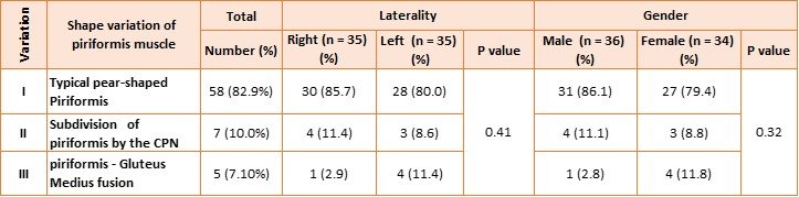
The typical pear-shaped morphology of the piriformis muscle (Variation I) (Figure 1D) was the most commonly observed pattern, identified in fifty-eight limbs (82.9%). Type II variation (Figure 1 E), in which the piriformis muscle was divided into two distinct parts by the common peroneal nerve , was observed in seven limbs (10.0%). Variation type III (Figure 1 F), characterized by fusion of the piriformis muscle with the gluteus medius muscle, was recognized in five limbs (7.1%).Comparison of the distribution of muscle variations showed no statistically significant differences regarding laterality (P = 0.41) and gender (P = 0.32) (Table 2).
Table 2: Shape variation of piriformis muscle according to laterality and gender.
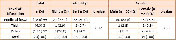
Regarding the level of sciatic nerve bifurcation, the popliteal fossa (Figure 1 A) represented the most common site of bifurcation, observed in 55 limbs (78.6%), whereas thigh bifurcation(Figure 1 B) in 3 limbs (4.3%) and pelvic bifurcation (Figure 1 C) was identified in 12 limbs (17.1%). Considering laterality and gender, no statistically significant differences were observed at the level of bifurcation (p > 0.05) (Table 3).
Table 3: Level of sciatic nerve bifurcation according to laterality and gender.
In (Table 4), the sciatic nerve–piriformis relationship was evaluated according to the Beaton and Anson classification. Type I configuration (Figure 2 A) represented the predominant anatomical pattern, identified in 55 limbs (78.6%). Type II variation (Figure 2 B) was observed in 7 limbs (10.0%), whereas Type III variation (Figure 2 C) was identified in 4 limbs (5.7%). Types IV, V, and VI (Figure 2 D, E & F) were uncommon findings. Beaton and Anson classification exhibited no statistically significant differences regarding laterality and gender (p > 0.05).
Table 4: Beaton and Anson classification patterns according to laterality and gender.
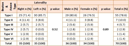
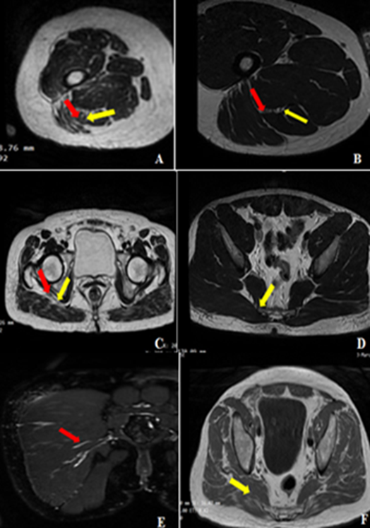
Fig. 1: Axial T2- weighted MRI images demonstrating levels of sciatic nerve bifurcation (A-C) at the upper border of popliteal fossa (A), upper thigh (B), sub gluteal region(C) . The red arrows represent the common peroneal nerve while the yellow arrow demonstrate the tibial nerve. Anatomical variation of Piriformis muscle (D-F): variation I (D) with typical pear-shaped morphology(yellow arrow). Variation II (E) in which the piriformis is divided into two parts with the common fibular nerve (red arrow) coursing between them. Variation III (F) demonstrates the union of the piriformis muscle with the gluteus medius muscle (yellow arrow).
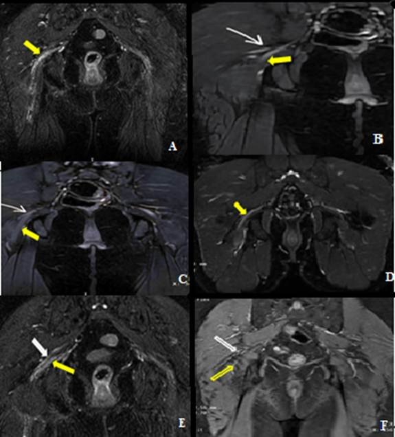
Fig. 2: Coronal T2-weighted MRI of the pelvis demonstrating the sciatic nerve–piriformis muscle relationship according to the Beaton and Anson classification. Type I (A): undivided sciatic nerve (yellow arrow) passing inferior to the piriformis muscle. Type II (B): common peroneal nerve (white arrow) passing through the piriformis muscle while the tibial nerve (yellow arrow) passes inferiorly.Type III (C): common peroneal nerve (White arrow) passing superior to the piriformis muscle with the tibial nerve (Yellow arrow) passing inferiorly.Type IV (D): undivided sciatic nerve (Yellow arrow) passing through the piriformis muscle. Type V (E): common peroneal nerve (White arrow) passing through the piriformis muscle while the tibial nerve ( Yellow arrow) passes superiorly. Type VI (F): undivided sciatic nerve (White arrow) passing superior to the piriformis muscle (Yellow arrow).
Discussion
In our study, the diameter and thickness of sciatic nerve at the sub gluteal level were 11.62 mm and 6.4 mm respectively. These findings support the elliptical morphology of the sciatic nerve, where the mediolateral diameter exceeds the anteroposterior thickness. Similar MRI-based measurements were reported by Rusu et al., who demonstrated mean sciatic nerve width and thickness values of approximately 10.4 mm and 5.5 mm, respectively [13]. The slightly higher values observed in our study may be attributed to differences in population characteristics and variations in imaging protocols.
Ultrasound-based studies report sciatic nerve width ranging from 12 to 20 mm and thickness between 4 and 8 mm at the sub gluteal level [14, 15] and the values obtained in our study fall within these ranges. Cadaveric studies, on the other hand, often demonstrate larger measurements, with sciatic nerve diameters reaching 15–20 mm [16].
Although cadaveric studies frequently report larger sciatic nerve dimensions, these differences may reflect inclusion of surrounding connective tissue, fixation-related changes, and methodological variation in measurement techniques [17].
The present MRI findings suggest that sex differences of sciatic nerve dimensions, with males demonstrating larger diameter (12.3 mm) and thickness (7.1 mm) than females (10.9 mm and 5.8 mm). Similar observations were reported by Karmakar et al. in their ultrasonographic evaluation, who suggested that anthropometric factors including sex may influence sciatic nerve dimensions during ultrasonographic evaluation of the subgluteal region (14). Rusu et al. also demonstrated that MRI provides reliable assessment of sciatic nerve morphology and noted that inter-individual variability may be partially related to sex and body composition differences [13]. The larger sciatic nerve dimensions observed in males in the current study may therefore reflect differences in body habitus, muscle mass, and lower limb morphology.
With respect to anatomical variation of Piriformis muscle, the typical pear-shaped muscle, identified by a single muscle belly, is the most frequent structure in anatomical studies. Similar findings have been reported in other studies, which in cadaveric specimens, demonstrated that the classic morphology dominates, around 67–70% of limbs examined and this pattern mirrors the role of piriformis muscle in hip external rotation and joint stability [18]. The two-headed piriformis which accounts for 10% of the study material in which the muscle is divided into two parts by the common fibular nerve. Bergman states that the two parts variation is related to high division of the sciatic nerve with the common fibular portion of the nerve exists between the two parts of Piriformis muscle [19].
A study on Polish cadavers reported that the two-headed piriformis was found in about 20% of cases, demonstrating that such a variation is one of the more common anatomical variants beyond the typical single-belly form. This foundation is of clinical significance since the changed relationship with the common fibular nerve can lead to nerve entrapment or affect the sciatic nerve course in the gluteal region The Less common variant, piriformis – gluteus medius fusion, was observed in 7.1% of our sample (5/70 limbs). The possible fusion of piriformis muscle with nearby muscles like Gluteus medius, superior gemellus and Obturator internus had been described by Bergman [19]. This finding was similar to the lower percentage found in in the Polish study. Literature reviews demonstrate that variations of the sciatic nerve’s relation to the Piriformis occur in over 10% of individuals (in our study approximately 17%) are comparable among populations without significant differences based on gender or laterality. Awareness of piriformis muscle variations can improve radiologic interpretation, influence operative techniques in the gluteal region, and advice clinicians when assessing unexplained sciatic pain [20].
The two portions of the sciatic nerve develop separately during early embryonic life and later join within a common connective tissue sheath to form a single nerve trunk. Thus, the point of cessation of this connective tissue wrapping determine the level of division of sciatic nerve into its main portions [21].
Hence, based on their embryonic development, it is possible that the bifurcation level of sciatic nerve may be in the gluteal region, the posterior thigh or at the apex of popliteal fossa. In this study, most sciatic nerve bifurcations occurred at the upper angle of the popliteal fossa (78.6%), whereas pelvic and thigh bifurcations represented less frequent anatomical patterns. These findings are consistent with previous anatomical and radiological studies identifying the popliteal region as the most common site of sciatic nerve division. Lakshmi Kumari et al. reported that approximately 69.2% of sciatic nerves bifurcated at the upper angle of the popliteal fossa, while pelvic and thigh bifurcations occurred less frequently [22]. Similarly, Grewal et al. demonstrated popliteal bifurcation in approximately 63.3% of examined limbs [23]. Comparable findings were also reported in South African cadaveric studies, where nearly 79.6% of sciatic nerves divided within the popliteal fossa [24]. The predominance of distal bifurcation observed in the present study therefore supports the concept that popliteal division represents the standard anatomical configuration in most individuals. High sciatic nerve bifurcation has important clinical implications because proximal division within the pelvis or thigh may contribute to failed sciatic nerve block, piriformis syndrome, and iatrogenic nerve injury during surgical procedures. Previous studies demonstrated that higher bifurcation patterns are associated with altered nerve pathways and incomplete regional anesthesia, particularly when division occurs proximal to the popliteal fossa [24].
In the present study, no statistically significant differences in bifurcation level were observed according to sex or laterality. Similar findings were reported by Schiariti et al. and Saleh et al., who demonstrated that sciatic nerve bifurcation patterns are generally independent of sex and side distribution [25,26].
Analysis of the sciatic nerve–piriformis relationship according to the Beaton and Anson classification demonstrated that Type I configuration represented the predominant anatomical pattern, whereas Types II and III were less common and Types IV–VI represented rare findings. These findings are consistent with previous cadaveric and imaging studies demonstrating that the undivided sciatic nerve passing inferior to the piriformis muscle is the most common anatomical arrangement. Large anatomical reviews and meta-analyses reported Type I prevalence ranging from 80% to 90% in different populations, whereas the remaining variants collectively account for a relatively small proportion of cases [27]. MRI and cadaveric studies similarly reported Type II and Type III variations in approximately 5–15% and 1–5% of examined limbs, respectively [28]. The clinical significance of Beaton and Anson variations lies in their relationship to sciatic nerve entrapment syndromes and regional anesthetic procedures. Abnormal pathways of the sciatic nerve relative to the piriformis muscle may predispose patients to piriformis syndrome and may alter the effectiveness of sciatic nerve block techniques. Therefore, accurate identification of these anatomical variants is important during MRI interpretation, posterior hip surgery, and image-guided interventions. In agreement with previous systematic reviews, the present study demonstrated no statistically significant association between Beaton and Anson classification and sex or laterality [27,29].
Conclusion
The present study demonstrated that the sciatic nerve diameter and thickness were significantly higher in males than females while no significant side-related differences were indicated. The typical pear-shaped Piriformis muscle is the most frequent structure and anatomical variations of the piriformis occur in approximately 17% without significant differences based on sex or laterality.
The level of sciatic nerve bifurcation in more than two third of our sample was at the apex of the popliteal fossa followed by pelvis and mid-thigh. According to Beaton and Anson classification Type 1 in 55 (78.6%) was the most common accounting more than three quarters of our sample followed in sequence by Type II in 3 limbs (10.0%) and Type III in 4 limbs (5.7%). Types IV, V, and VI espectively were recognized in two limb (2.9%). Type V 1 (1.4%), and Type VI 1 (1.4%).
Furthermore, level of sciatic nerve bifurcation and sciatic nerve-piriformis relationships showed no significant association with either gender or laterality. These findings highlight that sexual dimorphism may influence the size of sciatic nerve. In contrast, the anatomical patterns and relationships of the sciatic nerve remain comparable across sexes and sides.
Awareness of variations of the sciatic nerve’s relation to the piriformis muscle is important for anesthesiologists, Orthopedic surgeons, clinicians and radiologists during various clinical conditions.
Limitations: The present study has several limitations. First, the sample size was relatively small and derived from a single center, which may limit generalizability of the findings. Second, MRI-based morphometric assessment may be influenced by motion artifacts, partial volume effects, and image resolution limitations despite standardized imaging protocols. Third, the study focused primarily on anatomical and morphometric evaluation without correlation to clinical symptoms or electrophysiological findings.
Authors’ declaration: The project was approved by the Institutional Review Board of the Department of Anatomy, College of Medicine, University of Baghdad (Ref. No. 601; 30 June 2025).
Conflict of Insert: None
Authors’ contributions
Study conception and design: Malak; literature search: Mohammed and Malak; data analysis and interpretation: Mohammed and Malak; manuscript preparation, editing and review: Malak and Mohammed.
Cite this article: Mohammed Eimad Mohammed, Malak A. Al-Yawer. Sciatic Nerve Bifurcation Level and its Anatomical Relationship to the Piriformis Muscle: A Magnetic Resonance Imaging–Based Assessment in the Iraqi Population. Int J Anat Res. 2026;14(3):9680–9688. DOI: 10.16965/ijar.2026.206
References
- Standring S, ed. Gray's Anatomy: The Anatomical Basis of Clinical Practice. 41st ed. London: Elsevier; 2016.
- Moore KL, Dalley AF, Agur AMR. Clinically Oriented Anatomy. 8th ed. Philadelphia: Wolters Kluwer; 2018.
- Drake RL, Vogl W, Mitchell AWM. Gray's Anatomy for Students. 4th ed. Philadelphia: Elsevier; 2020.
- Netter FH. Atlas of Human Anatomy. 7th ed. Philadelphia: Elsevier; 2019.
- Hopayian K, Song F, Riera R, Sambandan S. The clinical features of piriformis syndrome: a systematic review. Eur Spine J. 2010;19(12):2095-2109. https://doi.org/10.1007/s00586-010-1504-9 PMid:20596735 PMCid:PMC2997212
- Pokorný D, Jahoda D, Veigl D, Pinskerová V, Sosna A. Topographic variations of the relationship of the sciatic nerve and piriformis muscle. Surg Radiol Anat. 2006;28(1):88-91. https://doi.org/10.1007/s00276-005-0056-x PMid:16311716
- Beaton LE, Anson BJ. The relation of the sciatic nerve and of its subdivisions to the piriformis muscle. Anat Rec. 1937;70:1-5. https://doi.org/10.1002/ar.1090700102
- Smoll NR. Variations of the piriformis and sciatic nerve with clinical consequence. Clin Anat. 2010;23(1):8-17. https://doi.org/10.1002/ca.20893 PMid:19998490
- Benzon HT, Raja SN, Molloy RE, Liu SS, Fishman SM. Essentials of Pain Medicine. 4th ed. Philadelphia: Elsevier; 2018.
- Tubbs RS, Salter EG, Wellons JC, Blount JP, Oakes WJ. Anatomical landmarks for the sciatic nerve in the gluteal region. J Neurosurg. 2005;102(2):193-195. https://doi.org/10.3171/spi.2005.2.3.0335 PMid:15796359
- Chhabra A, Faridian-Aragh N. High-resolution MR neurography of sciatic neuropathy. AJR Am J Roentgenol. 2012;198(3):W357-W364. https://doi.org/10.2214/AJR.11.6981 PMid:22451573
- Beaton LE, Anson BJ. The relation of the sciatic nerve and of its subdivisions to the piriformis muscle. Anat Rec. 1937;70:1-5. https://doi.org/10.1002/ar.1090700102
- Rusu GM, Curcean AD, Dudea SM. Ultrasound and magnetic resonance imaging evaluation of the femoral and sciatic nerves: a study of healthy volunteers. Med Ultrason. 2020;22(4):1-7. https://doi.org/10.15386/mpr-1629 PMid:33225261 PMCid:PMC7664720
- Karmakar MK, Shariat AN, Pangthipampai P, Chen J. Sonoanatomy of the sciatic nerve in the subgluteal region. Br J Anaesth. 2007;98(3):390-395. https://doi.org/10.1093/bja/ael364 PMid:17307781
- Vloka JD, Hadzić A, Lesser JB, Kitain E, Geatz H, April E, Thys DM. The division of the sciatic nerve in the popliteal fossa: anatomical implications for regional anesthesia. Anesth Analg. 2001;92(1):215-217 https://doi.org/10.1097/00000539-200101000-00041 PMid:11133630
- Standring S, ed. Gray's Anatomy: The Anatomical Basis of Clinical Practice. 41st ed. Philadelphia: Elsevier; 2016.
- Ma, C., Wang, X., & Li, J. Assessment in location of sciatic nerve between the ischial tuberosity and the greater trochanter of the femur: A cadaveric study. Heliyon, 2024;10(1):e23751. https://doi.org/10.1016/j.heliyon.2023.e23751 PMid:38192877 PMCid:PMC10772174
- Haładaj R, Pingot M, Polguj M, Wysiadecki G, Topol M. Anthropometric Study of the Piriformis Muscle and Sciatic Nerve: A Morphological Analysis in a Polish Population. Med Sci Monit. 2015 Dec 2;21:3760-3768. https://doi.org/10.12659/MSM.894353 PMid:26629744 PMCid:PMC4671451
- Bergman RA, Thompson SA, Aff AK, Saadeh FA: Compendium of human anatomic variation: text, atlas, and world literature. Baltimore and Munich: Urban & Schwarzenberg, 1988.
- Poutoglidou F, Piagkou M, Totlis T, Tzika M, Natsis K. Sciatic Nerve Variants and the Piriformis Muscle: A Systematic Review and Meta-Analysis. Cureus. 2020 Nov 17;12(11):e11531. https://doi.org/10.7759/cureus.11531
- Babinski MA, Machado FA, Costa WS. A rare variation in the high division of the sciatic nerve surrounding the superior gemellus muscle. European journal of Morphology. 2003;41(1):41-2. https://doi.org/10.1076/ejom.41.1.41.28099 PMid:15121548 PMCid:PMC12457634
- Lakshmi Kumari K, Sushma M, Raja A, Asha Latha D. Anatomical study on sciatic nerve variations in Andhra Pradesh, India. International Journal of Research in Medical Sciences. 2019 Aug;7(8). https://doi.org/10.18203/2320-6012.ijrms20193399
- Grewal H, Singla RK, Singh R, Singla M. Different levels of bifurcation of sciatic nerve: A novel classification based on a cadaveric study in Indian population. International Journal of Anatomy and Research. 2016;4(3):2743-2749. https://doi.org/10.16965/ijar.2016.323
- Kegoye, E. S., Ojewale, A. O., Ezekiel, W., et al. Morphologic and morphometric bilateral analysis and sexual dimorphism in sciatic nerves of adult cadaveric specimens in Uganda. BMC Musculoskeletal Disorders, 2025;26(1):422. https://doi.org/10.1186/s12891-025-08641-9 PMid:40301790 PMCid:PMC12039151
- Schiarite, L., Calvo, M., Maggi, G., Abad, A., & Gilsanz, F. Location of the sciatic nerve at the popliteal fossa in the adult population: Relationship with gender and anthropometric data studied. Implications for daily practice. Revista Española de Anestesiología y Reanimación, 2015;62(10):565-569. https://doi.org/10.1016/j.redar.2015.02.012 PMid:25896734
- Saleh, H. A., El-Fark, M., & Abdel-Hamid, G. A. Anatomical variation of sciatic nerve division in the popliteal fossa and its implication in popliteal nerve blockade. Folia Morphologica, 2009;68(4):256-259.
- Poutoglidou F, Piagkou M, Totlis T, Tzika M, Natsis K. Sciatic Nerve Variants and the Piriformis Muscle: A Systematic Review and Meta-Analysis. Cureus. 2020 Nov 17;12(11):e11531. https://doi.org/10.7759/cureus.11531
- Frideriki Poutoglidou, Maria Piagkou, Trifon Totlis, Maria Tzika, Konstantinos Natsis, Sciatic Nerve Variants and the Piriformis Muscle: A Systematic Review and Meta-Analysis, Cureus. 2020 Nov 17;12(11):e11531, https://doi.org/10.7759/cureus.11531
- Marchese CR, Graves AL, Pautler BJ, Dye D, Creamer BA, Dennis JF. Beaton and Anson Type A Classification of the Sciatic Nerve and Piriformis Complex: Clinical Considerations for Sex and Laterality. Anatomia. 2024 Aug 21;3(3):182-191. https://doi.org/10.3390/anatomia3030014.


