
Chromosomal Mosaicism at a Tertiary Care Center in Central India
Manisha B.Sinha · Int J Anat Res 2026;14(3):9654–9660
1 Professor, Lab of Molecular and Cytogenetics, Department of Anatomy, All India Institute of Medical Science Raipur C. G, India.
Corresponding Author: Dr.Manisha B.Sinha, Professor, Lab of Molecular and Cytogenetics, Department of Anatomy, All India Institute of Medical Science Raipur, C. G, India. · manishabsinha@gmail.com
Abstract
Background
Chromosomal mosaicism refers to the presence of two or more genetically distinct cell lines within an individual derived from a single zygote. The clinical expression varies widely depending on the proportion and distribution of abnormal cells. Objective: To evaluate the spectrum, clinical presentation, and outcomes of chromosomal mosaicism in individuals with suspected chromosomal abnormalities at a tertiary care center.
Methods
This observational study included 308 individuals with suspected chromosomal disorders evaluated between January 2024 and March 2026. Peripheral blood samples were collected and cultured using standard cytogenetic techniques. Karyotyping was performed using G-banding.
Results
Chromosomal mosaicism was identified in 9 cases (2.9%). The majority involved sex chromosome mosaicism, predominantly mosaic Turner syndrome variants. Common clinical indications included primary amenorrhea and short stature. Structural abnormalities of the X chromosome were also observed. One case of mosaic trisomy 21 and one case of 45,X/46,XY mosaicism were identified. Mosaic cases generally exhibited milder or incomplete phenotypic features compared to classical syndromic presentations.
Conclusion
Chromosomal mosaicism is associated with variable and often attenuated clinical manifestations. Low-level mosaicism tends to correlate with better clinical outcomes and survival. Early detection is important for appropriate management and genetic counselling. Mosaic cases, particularly when identified prenatally, may have favourable prognostic implications.
Background
Chromosomal mosaicism is defined as the presence of two or more genetically distinct cell populations within a single individual arising from a single fertilised egg. This phenomenon results from postzygotic genetic alterations and contributes to a wide range of clinical conditions, spanning from localised developmental anomalies to systemic genetic disorders and malignancies.
The phenotypic expression of mosaicism depends on several factors, including the timing of the mutational event during embryogenesis, the proportion of abnormal cells, and their distribution across tissues. Mosaicism may involve somatic cells, germ cells, or both, and is broadly classified into somatic mosaicism, germline mosaicism, and combined (gonosomal) mosaicism.
Clinical features of mosaicism frequently manifest as asymmetrical tissue development (e.g., focal cortical dysplasia or segmental hypertrophy), patchy cutaneous pigmentation along lines of Blaschko, and localized vascular malformations
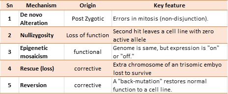
Mechanisms causing mosaicism (Table 1)
- De novo genetic alteration:
- Epigenetic Mosaicism:
- Mosaic Nullizygosity:
- Rescue mechanisms:
- Postzygotic loss
- Spontaneous reversion
- Postzygotic loss
Table 1 represents the different mechanisms of mosaicism.
Table 1: Showing the mechanism for different alterations.
Mosaic manifestations of Mendelian disorders: it is sub-grouped as
- Mosaicism for lethal mutations exists only in mosaic forms. Death occurs in a few years of life. Because it is lethal, it does not transfer to offspring.
- Mosaicism for mutations known in autosomal -dominant/X linked recessive disorders: Occurs in Neurofibromatosis type 1, Neurofibromatosis type 2 as somatic mosaicism 25.33% and 6.5% respectively [1]. Severity depends on the time of mutation at the postzygotic stage. Clinical manifestations presented as an atypical and disseminated form. Sometimes they present atypically, away from the expected manifestations of this disorder.
In these, parents are with somatic mosaicism.
- Rare mosaicism that happened as aggravation of an in-segmental area phenotype: this type occurs in a subpopulation of precursor cells having a germline mutation.
Chromosomal mosaicism in fetuses leads to abortions. Constitutional gain or loss of a few chromosomes, such as 13, 18, 21, and X, can occur in mosaic form [2]. Mosaicism in partial deletion of X and normal X is also commonly observed in primary amenorrhea and infertility cases. These people have better cognitive function. They have a milder phenotype. Other additional chromosomal aneuploidies occur only in mosaic form, such as chromosomes 8, 9, 14, 17, and 22. They are not viable for life if present in constitutional form. The presentation of the sign is milder. It totally depends on the number of cells in the mainstream. However, the correlation between the number of mosaic cells and clinical features has not yet been established. The presence of patchy pigmentation will suggest mosaicism.
Various parameters, such as maternal age [3], consanguinity [4,5], socioeconomic status [6,7], ethnicity [8], play a role in the development of chromosomal abnormalities, including mosaicism.
Chen et al. in 2022 found mosaic trisomy 18 at amniocentesis at 18 weeks. Mosaicism was 30%, 47,XX,+18[6]/46,XX[17] in that case by karyotyping. Chromosomal array (comparative genomic hybridization- aCGH) on uncultured amniotic fluid cells showed 45% mosaicism. Later in the pregnancy, the mosaicism percentage has decreased. In the same 1½-month-old child, the blood karyotype showed 14% mosaicism for trisomy 18, and the buccal mucosa karyotype was 2% [9]. This implies that the lowering mosaic proportion over time is explained by somatic selection favoring normal cell lines in quickly dividing tissues (such as blood and buccal mucosa), which lowers morbidity with mosaicism.In a study of cohort 500, 12 cases were found to be mosaic [10]. Three proband and nine parents were received a diagnosis of mosaicism.
Identifying mosacism in a proband and then to a family member or a parent is important. These have recurrence risk. De novo pathogenic variants have a recurrence risk̴ ̴ 1%. This is an empirical risk estimate that accounts for the rare potential of germline parental mosaicism; nonetheless, discovery of the same pathogenic mutation in a parent can greatly raise the recurrence risk (up to 50%). De novo pathogenic variant in postzygotic.
Advances in cytogenetic and molecular techniques have improved the detection of mosaicism; however, conventional karyotyping remains a widely accessible and essential diagnostic tool in resource-limited settings.
The present study aims to evaluate the frequency, clinical spectrum, and cytogenetic patterns of chromosomal mosaicism in individuals presenting with suspected chromosomal abnormalities at a tertiary care center, and to correlate these findings with clinical presentation and outcomes.
Materials and methods:
After obtaining the institute’s ethical clearance, the study was conducted in a cohort of 308 participants with suspected chromosomal disorders from Jan 2024 to March 2026. A blood sample was collected in a heparinized bottle. After taking consent and a demographic profile, the sample was cultured. After 70 hours of culturing, harvesting, and slide preparation, the process was completed. Giemsa stain was used to obtain bands. Karyotype images were taken under a microscope (Carl Zeiss, Oberkochen, Germany). In suspected cases where over two cell lines were observed, more than fifty metaphases were examined using the automated system (300-850 band levels of resolution).
Result:
Cases of Mosaicism: There were nine cases of chromosomal mosaicism. Four patients were visited at the OPD with a primary complaint of amenorrhea. Three girls had the primary complaint of short stature. One baby had syndromic features. One Child had the primary complaint of micropenis.
Case 1:
A 4-year-old female child, weight 12.81kg, height 90.6cm, head circumference 45cm. Height-for-Age was -2.8SD, which indicates the child is significantly shorter than average. A value below -2.0SD (Standard deviation) is considered short stature. Head Circumference-for-Age was -3SD, showing a head circumference significantly smaller than average for their age. W/A (Weight-for-Age) = -1.7SD. The child’s weight was below average for their age, but less severely impacted than their height. W/H (Weight-for-Height) = 0.0SD: This is a positive sign. Child’s weight is perfectly proportional to their current height. The iron profile was absolutely normal. The vitamin D 25-hydroxy serum level was lower (15.7). Other blood parameters were within normal limits. Karyotype of the case was mos 46,X,del(X)(q26)[14]/46,XX[46]. The most prominent manifestation in turner syndrome is short stature, which is supposed to result from haploinsufficiency of genes and/or loss of distal regulatory elements located beyond Xq26 that are required for normal skeletal growth and development. The severity of the phenotype depends on the extent of the deleted segment and the degree of mosaicism.
Case 2:
A 16-year-old female visited with her parents with a primary complaint ofamenorrhea. Other signs and symptoms included a high-arched palate, widely spaced nipples, cubitus valgus, B/L short 4th fingers, short stature, underdeveloped secondary sexual characteristics, and an aortic murmur. On USG examination, a hypoplastic uterus (3×1.2×0.4cm3) and ovaries (Right 1.7×0.8cm2, left 1.3×0.5cm2) were observed. Hormonal essay revealed LH- 42.49mIU/ml(high), FSH – 193.57mIU/ml (high), Testosteron- 7ng/dl(borderline), Prolactine- 9.49ng/ml(normal). Karyotype was mos,45,X[55]/46,X,+mar[5] (figure 1). The treatment plan would depend on whether the marker carries the SRY gene. This was a complex scenario. MLPA ( using P095-A4 ANEUPLOIDY Kit) showed the absence of the SRY gene.
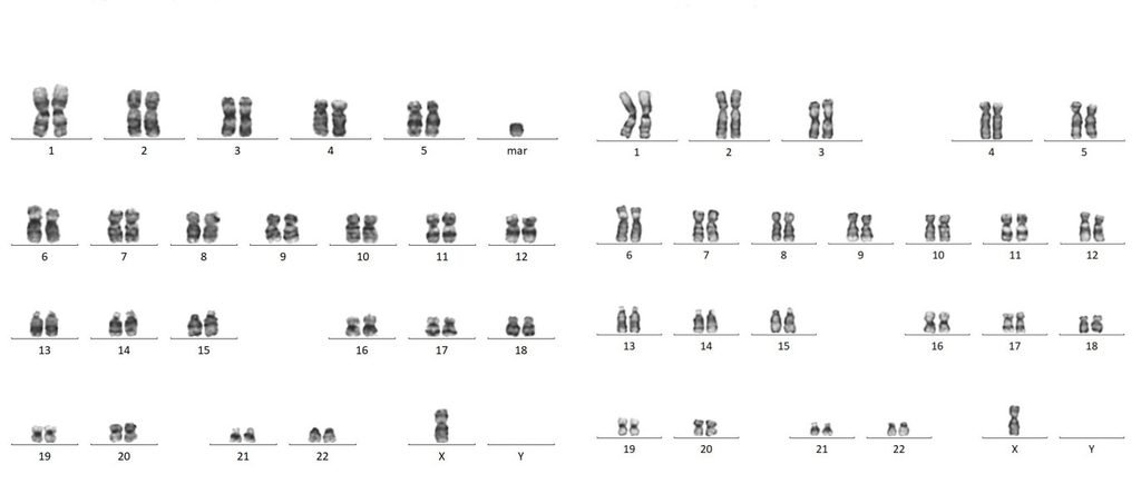
Fig. 1: Karyotype of case1- 46,XO,+mar/ 45,XO.
Case 3:
One of these was a 25-year-old female who complained of primary amenorrhea. On Tanner staging, breast development was grade III, and pubic hair development was grade II to III.
USG findings showed an infantile uterus with fewer follicles seen in both ovaries. Karyotype of the patient was Mos 45,XO[4]/46,XX[48].
Case 4:
A 25-year-old female came with a complaint of amenorrhoea. On USG of the pelvis, a hypoplastic uterus of 4.5×2.5×1.6cm3, volume 10.6 cc, and endometrial thickness 5.7mm was found. The right and left ovaries were normal, and a 2×1.7cm2 functional cyst was in the left ovary. TSH level (2.39µIU/ml), LH level (4.51mIU/ml), and FSH level (3.21 mIU/ml) were normal. Karyotype of case was mos46,XX,del(X)(q27-28)[25]/46,XX[48].
Case 5:
Another case of primary amenorrhea, age 25 years, visited the OPD; on Tanner staging, breast development was grade III, pubic hair development was grade III. On USG and MRI of the pelvis, an Infantile hypoplastic uterus (2.0 x 1.0 x 2.2cm3) with fewer follicles was seen in both ovaries. Levels of hormone were TSH level 1.21mIU/ml(normal), FSH – 53.6mIU/ml(high),LH-29.1mIU/ml(high), prolactin 5.66ng/ml. Karyotype was Mos 46,XX,del(X)(q26-ter)[4]/46,XX[46].
Case 6:
A case of micropenis with bilateral palpable testes was seen in OPD. USG abdomen showed normal features. Testosterone level – 18.11ng/dl (low), TSH- 3.38mIU/ml (normal). The patient’s karyotype showed mosaicism, mos,45,X[20]/46,XY[30]. This patient was under regular follow-up for testicular carcinoma. This type of patient has dysgenetic gonads. Testosterone replacement therapy has been initiated under pediatric endocrine supervision to induce puberty and promote penile growth.
Case 7:
Male child of 2425g birth weight, delivered by LSCS at 33 weeks of gestation with G3P1L1A1; the age of the mother was 29 years at the birth of the baby. She had pregnancy-induced hypothyroidism. The child had bilateral undescended testes. The X-ray showed a double Bubble appearance. On USG examination, duodenal Atresia Type 1, non-rotation of the large bowel, and dextrocardia were found. The operation done was an exploratory laparotomy and a Kimora duodeno-duodenostomy. Karyotype of the baby was mos,47, XY,+21[20]/46,XY[30].
Case 8:
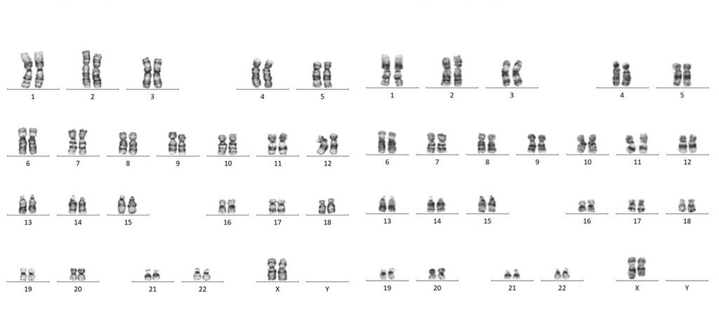
A seven-year-old female child, weighing 15.3kg and measuring 107cm in height, presented with an upper respiratory tract infection. She visited the OPD with the primary complaint of short stature, restricted growth, and patchy hyperpigmentation on the elbow. Her upper segment of body: lower segment of body was 57:59, 0.96:1. Karyotype was mos46,XX,del(X)(q26)[15]/46,XX[39] (Figure 2). The physical embryology of somatic cell lineage migration is vividly highlighted by the hyperpigmentation that followed Blaschkan lines or developmental whorls.
Fig. 2: Karyotype of case 2-46,XX/46,XX,del(X)(q26).
Case 9:
A nine-year-old girl with height 111.4cm, weight 25kg, serum iron and 25-hydroxy vitamin D were low, and TSH was increased. Chest X-ray bilateral clear. Treatment started with thyroxine. Karyotype was mos 45,XO[25]/46,XO,+mar[25]. MLPA report (using P095-A4 ANEUPLOIDY Kit) of the case showed absence of the SRY gene. This patient was also for follow-up.
Discussion
In this single-center observational cohort of 308 individuals evaluated for suspected chromosomal abnormalities, chromosomal mosaicism was identified in 8 cases (2.6%). This frequency is broadly consistent with prior genomic cohort data, such as the study by Cook CB et al., which reported mosaicism in 12 of 500 cases (~2.4%), supporting that mosaicism represents a small but clinically significant subset in diagnostic genetics.
A key observation in our series is the predominance of sex chromosome mosaicism, particularly mosaic monosomy X and its structural variants. Most patients presented with classical but attenuated Turner phenotype features, including short stature, primary amenorrhea, and variable gonadal dysgenesis. However, compared with non-mosaic counterparts, phenotypic expression was milder and heterogeneous. This aligns with established concepts that the proportion and tissue distribution of abnormal cell lines critically influence clinical severity, although precise genotype–phenotype correlations remain inconsistent.
Notably, structural mosaic abnormalities of the X chromosome (e.g., del(Xq)) were associated with endocrine dysfunction and reproductive anomalies, even in the presence of a significant proportion of normal cell lines. It was observed that 6-12 % of mosaic Turner cases with the Y chromosome element [11]. Prophylactic gonadectomy should be done as they have an increased risk of developing gonadoblastoma [12], a precursor to dygerminoma.
The detection of marker chromosomes and the need for further molecular characterization (e.g., SRY status by MLPA) highlight the importance of adjunct techniques beyond conventional karyotyping for risk stratification, particularly regarding gonadoblastoma in cases with Y chromosomal material.
The inclusion of mosaic 45,X/46,XY disorder of sex development (DSD) further illustrates the phenotypic variability inherent to mosaicism. These individuals require longitudinal surveillance due to risks of gonadal malignancy and endocrine dysfunction. Clinical management of 45,X/46,XY DSD requires a multidisciplinary approach because the phenotype is highly variable and depends on the degree of testicular development and fetal hormone production. Management typically involves pediatric endocrinologists for endocrine evaluation and long-term hormonal care, pediatric surgeons or pediatric urologists for assessment and management of gonadal and genital anatomy when indicated, and genetic counsellors to explain the diagnosis, recurrence risk, and implications for affected individuals and their families. Care should be individualized according to the patient’s phenotype, gonadal function, internal reproductive anatomy, and clinical needs, particularly because ductal development may range from müllerian structure retention to wolffian duct stabilization depending on fetal testicular function.
Autosomal mosaicism was less frequent but clinically relevant. One case of mosaic trisomy 21 demonstrated variable growth and developmental profiles, with presenting multiple congenital anomalies. These findings support prior observations that mosaic trisomies, especially involving chromosome 21, are compatible with survival and often associated with a milder phenotype compared to full trisomy.
Importantly, our data reinforce that mosaicism may present with subtle or atypical clinical features, such as patchy pigmentation or isolated growth abnormalities, necessitating a high index of suspicion. The variability in clinical expression observed across cases reflects the timing of the postzygotic event and the distribution of the mosaic cell line across tissues.
Conclusion
Chromosomal mosaicism constitutes a clinically significant but relatively infrequent finding. In current study we found 2.9% (9/308) cases in a setup of cytogenetic laboratory at tertiary care center. Among individuals with suspected chromosomal disorders. In our cohort, mosaic cases were predominantly associated with sex chromosome abnormalities, particularly mosaic Turner variants.
These cases typically exhibit attenuated or incomplete phenotypic manifestations compared to their non-mosaic counterparts, contributing to improved survival and, in some instances, near-normal functional outcomes. The presence of low-level mosaicism appears to correlate with a more favorable prognosis, although clinical variability remains substantial.
Early detection of mosaicism is essential for appropriate clinical management, genetic counseling, and risk assessment, particularly in relation to reproductive outcomes and malignancy risk. Our findings support the notion that mosaic chromosomal abnormalities are often compatible with life and may justify a more conservative approach in prenatal settings, provided detailed evaluation and counseling are undertaken.
Declaration: There is no conflict of interest among author/s.
Author contribution:
Manisha B Sinha: Data estimation, Resources, Data acquisition, Data analysis, Statistical analysis, writing- Original draft, Project administration, funding acquisition.
Acknowledgment
The author thanks Nursing Officer Ms. Renu and Lab Technician Mr. Chetan for their help throughout this study. The author also extends our regards to the family members who continue to participate in a better understanding
Cite this article: Manisha B.Sinha. Chromosomal Mosaicism at a Tertiary Care Center in Central India. Int J Anat Res. 2026;14(3):9654–9660. DOI: 10.16965/ijar.2026.192
References
- Ruggieri and Huson. The clinical and diagnostic implications of mosaicism in neurofibromatosis. Neurology. 2001; 156(11): 1433-43. https://doi.org/10.1212/WNL.56.11.1433 PMid:11409413
- Nagaoka SI, Hassold TJ, Hunt PA. Human aneuploidy: mechanisms and new insights into an age-old problem. Nature Reviews Genetics. 2012;13(7):493-504. https://doi.org/10.1038/nrg3245 PMid:22705668 PMCid:PMC3551553
- Stene J, Fischer G, Stene E, Mikkelsen M, Petersen E. Paternal age effect in Down's syndrome. Ann Hum Genet. 1977; 40: 299-306. https://doi.org/10.1111/j.1469-1809.1977.tb00194.x PMid:139837
- Puri RK, Khanna KK, Bhargava I, Balakrishnan S. Role of consanguinity in chromosomal syndromes. Indian J Med Res. 1977 Jun;65(6):859-64. PMID: 604263.
- Ray A, Oliver TR, Halder P, Pal U, Sarkar S, Dutta S, Ghosh S. Risk of Down syndrome birth: Consanguineous marriage is associated with maternal meiosis-II nondisjunction at younger age and without any detectable recombination error. Am J Med Genet. 2018;176: 2342-2349. https://doi.org/10.1002/ajmg.a.40511 PMid:30240118
- Hunter, J.E.; Allen, E.G.; Shin, M.; Bean, LJ Correa A, Druschel C Hobbs CA, O'Leary LA, Romitti, PA, Royle MH et al. The association of low socioeconomic status and the risk of having a child with Down syndrome: A report from the National Down Syndrome Project. Genet Med. 2013; 15: 698-705. https://doi.org/10.1038/gim.2013.34 PMid:23558253
- Vrijheid M, Dolk H, Stone D, Abramsky L, Alberman E, Scott JE. Socioeconomic inequalities in risk of congenital anomaly. Arch Dis Child. 2000; 82: 349-532. https://doi.org/10.1136/adc.82.5.349 PMid:10799420 PMCid:PMC1718336
- Kovaleva NV, Cotter PD, Mosaicism for Autosomal Trisomies: A Comprehensive Analysis of 1266 Published Cases Focusing on Maternal Age and Reproductive History. Genes. 2024;13(6):778. https://doi.org/10.3390/genes15060778 PMid:38927714 PMCid:PMC11202781
- Chen CP, Hsu TY, Tsai CC, Chern SR, Chen SW, Wu FT, Wu PS, Lee CC, Chen LF, Pan CW, Wang W. Mosaic trisomy 18 at amniocentesis associated with a favorable fetal outcome in a pregnancy. Taiwan J Obstet Gynecol. 2022; 61(4): 690-694. https://doi.org/10.1016/j.tjog.2022.05.006 PMid:35779923
- Cook CB, Armstrong L, Boerkoel CF, Clarke LA, du Souich C, Demos MK, Gibson WT, Gill H, Lopez E, Patel MS, Somatic mosaicism detected by genome-wide sequencing in 500 parent-child trios with suspected genetic disease: clinical and genetic counseling implications. Cold Spring Harbor Molecular Case Studies. 2021;7(6): a006125. https://doi.org/10.1101/mcs.a006125 PMid:34697084 PMCid:PMC8751411
- Bispo AV, Burégio-Frota P, Oliveira dos Santos L, Leal GF, Duarte AR, Araújo J, Cavalcante da Silva V, Muniz MT, Liehr T, Santos N. Y chromosome in Turner syndrome: detection of hidden mosaicism and the report of a rare X;Y translocation case. Reprod Fertil Dev. 2014 Oct;26(8):1176-82. https://doi.org/10.1071/RD13207 PMid:25294360
- Shen W, Li Y. Gonadoblastoma in Turner syndrome with puberty delay: A case report and literature review. Mol Genet Genomic Med. 2023; 11(12): e2300. https://doi.org/10.1002/mgg3.2300 PMid:37822273 PMCid:PMC10724510


