
Mapping the Arterial Architecture of the Hand: A Cadaveric Study Using Vascular Silicone Gel Injection
Pranup Roshan Quadras , Susie Jeyalyn David , Prima Swetha D’Souza , Dane Chandy · Int J Anat Res 2026;14(3):9646–9653
1 Associate Professor, Department of Anatomy, AJ Institute of Medical Sciences, Mangalore, Karnataka, India.
2 Associate Professor, Department of Anatomy, Sree Mookambika Institute of Medical Sciences, Kulasekharam, Kanyakumari District, Tamilnadu, India.
3 Assistant Professor, Department of Anatomy, Nitte(Deemed to be University) KS Hegde Medical Academy (KSHEMA), Mangalore, Karnataka, India.
4 Associate Professor, Department of Anatomy, Father Muller Medical College, Mangalore, Karnataka, India.
Corresponding Author: Dr. Susie Jeyalyn David, Associate Professor, Department of Anatomy, Sree Mookambika Institute of Medical Sciences, Kulasekharam, Kanyakumari District, Tamilnadu, India. · drsuslyn@gmail.com
Abstract
Background
The blood supply to the hand is provided by superficial palmar arch (SPA) and deep palmar arch (DPA). The knowledge of the arterial pattern is essential not only to anatomists, but also to surgeons, to carry out successful hand surgeries and to harvest radial artery (RA) as a graft for bypass surgery. Aims and Objectives: 1. To study the major and smaller branches which are not seen during routine dissection using vascular dye injection technique. 2. To study the variation of branching pattern of these arteries and their relevance in terms of clinical implications.
Methods
20 hands were selected randomly from 10 human cadavers. During embalming of the bodies, heparin was mixed in the embalming fluid to ensure that the vessels were cleared of clots and the lumen was patent. A flowable silicone compound (RTV116) was injected through the axillary artery and after one week, the hand specimens were dissected to expose the arches.
Results
20 hands were selected randomly from 10 human cadavers. During embalming of the bodies, heparin was mixed in the embalming fluid to ensure that the vessels were cleared of clots and the lumen was patent. A flowable silicone compound (RTV116) was injected through the axillary artery and after one week, the hand specimens were dissected to expose the arches.
Conclusion
The use of RTV 116 via vascular injection technique was beneficial to visualize the arch along with its minute branches and carry out meticulous dissection. Few variations and asymmetry with regards to SPA were observed.
INTRODUCTION:
The hand is the most complex part of the human body, involved in skilled movements and fine handling of objects. The human hand can be easily modulated to carry out certain compound movements with great precision, as per our need. Complex skilled movements require the hand to be placed and held in various anatomical positions. As a result, the entire framework of the human hand is supplied with an abundance of highly branched and anastomosing arteries which provides adequate blood supply.
The earliest evidence of anatomical study on the vascular system of the hand dates back to the 16th century. Andreas Versalius, in his publication, ‘De Humani Corporis Fabrica’, described the arterial supply of the hand. However, in his study, he did not specifically mention the presence of superficial and deep palmar arches. In 1961, Coleman and Anson [1] reported the presence of superficial and deep palmar arches in a study carried out on 650 cadavers. Furthermore, this was also later confirmed by Gellman et al, wherein they stated that both arterial arches were present in all their cases [2].
The arterial supply of the hand is by both, ulnar and radial arteries. The terminal parts of the ulnar and radial arteries on reaching the palmar aspect of the hand anastomose with each other to form superficial and deep palmar arches. The superficial branch remains to be a direct continuation of ulnar artery and forms the main contribution of the superficial palmar arch (SPA). The arch is eventually completed by one of the branches of the radial artery. The superficial palmar arch then gives four branches. One of which happens to be a proper branch to the little finger medially, and the other three are common digital arteries. These common digital arteries then further subdivide in pairs along the web spaces to form the proper palmar digital arteries. These all put together supply the medial three and a half fingers of the hand [3].
The human hand is supplied mainly by the superficial palmar arch (SPA) and deep palmar arch (DPA). The SPA forms a vascular arcade across the palm. The SPA can be classified into “complete” and “incomplete” SPA, depending on the presence or absence of anastomosis between the vessels forming the SPA [2].The most classical form of SPA is the “complete radio-ulnar” type, which is usually formed between the superficial branch of ulnar artery and is completed by a branch of the radial artery [1].
Anatomical variations have been observed with SPA and its contributing arteries which have been reported in literature [4,5]. Various hypotheses have been made to explain the occurrence of such variations.
It has been suggested that certain regulatory factors like hemodynamic forces, availability of oxygen and nutrient requirements during angiogenesis play a role in determining the arterial pattern at the time of foetal development. It should be also noted that the arterial patterns are highly reproducible and therefore genetic basis for the occurrence of such variations should not be undermined [6].
With the advent of microvascular surgeries for revascularization of the hand, replantations, grafting of various parts of the limbs and correcting deformities, knowledge of the arterial variations of the hand is essential for carrying out successful hand surgeries. More recently, radial arterial grafts are being used in bypass surgeries. The presence of a complete palmar arch and its proper identification are necessary in order to extract the radial artery [7].
A detailed study of the arterial supply is needed in order to assist not only surgeons, but also anatomists, who during dissection can be prepared to encounter unusual vascular patterns in the hand.
Although there are many previous studies on SPA, they are most often done on dissected embalmed cadavers which make it hard to distinguish the minute branches of SPA. To bridge the gap, the present study aims in executing a meticulous dissection to find out the frequency of variations in SPA patterns using coloured silicone gel injection technique.
Aims And Objectives:
- The aim of the study is not only to study the major branches but also the smaller branches which are not seen during routine dissection using vascular injection technique.
- To study the variation of branching pattern of these arteries and their relevance in terms of clinical implications
MATERIALS:
- Dissection kit (Fig 1)
- RTV 116 (flowable silicone dye) (Fig 2)
- Thread to ligate the cut ends of the artery
- Syringes
- Cannula
- Hand held digital camera
- Magnifier
- Light source
- Display signs
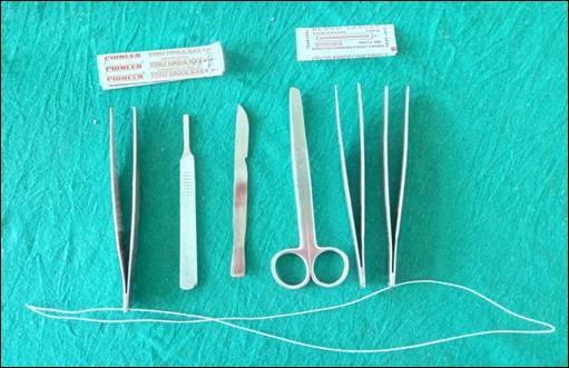
Fig. 1: Dissection Instruments.
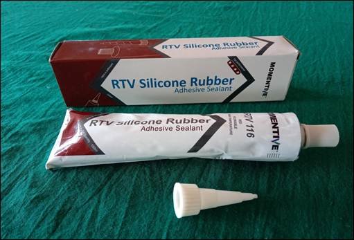
Fig. 2: RTV 116 (flowable silicone dye).
METHODS:
Study Design: Cross sectional study.
Sample Size: 20 hands were selected from randomly chosen 10 human cadavers for the study.
Inclusion Criteria: Hands from human cadavers, both male and female cadavers
Exclusion Criteria: Gross pathology, if any, was excluded. (Ex: Damaged specimen, missing fingers, congenital anomalies, etc)
The study was conducted in the Department of Anatomy, K.S Hegde Medical Academy. Ethical clearance was obtained from the Institutional Ethical Committee before the start of the study.
The upper limbs chosen in this study, for the arterial supply of the hand, were taken from well embalmed bodies. Separate vial of Heparin 25,000 IU was mixed in the embalming fluid (10% formalin with glycerine) to get the vessels cleared of any blood clot remnants and keep the lumen patent. This procedure helps in making the lumen of the blood vessels more accessible for vascular injection. These well embalmed bodies were placed in formalin filled storage tanks in our preservative room which was attached to the dissection hall. These tanks are filled with 10% formalin with glycerine and thymic granules. This preservative mixture was changed periodically.
Dissection pattern followed was as prescribed by Cunningham’s manual of dissection [8]. The axillary artery distal to the pectoralis minor muscle was identified during dissection of the axilla. A small transverse nick was made and the cannula was inserted. A flowable silicone compound (RTV116), available commercially, is red in colour. It was taken in a 50 ml syringe and then fixed into the cannula for injection into the axillary artery. About 50 ml of RTV116 was injected. After the dye was injected, the proximal end of axillary artery was ligated and was kept for seven days. It is known that the silicone compound (RTV116) when exposed to air starts hardening. The hardening is usually completed between three to six days. After seven days, the hand was dissected. The ulnar artery and radial artery were isolated and its branches studied. Since the silicone compound (RTV116) is red in colour, even the minute branches which were not visible to the naked eye were seen clearly as it was filled with the red colour dye. This facilitated dissection and all the major branches and minute branches of the arteries supplying the hand could be visualized and studied. The variations seen during dissection were recorded. Photographs were taken for documentation.
Collection and Recording of data
- Data analysis was done using Microsoft Excel.
- Qualitative data were represented as frequency and percentage.
- The results were compared with previous studies and variations of superficial palmar arch were plotted in tables.
RESULTS:
Patterns of Superficial Palmar Arch:
In this study, the superficial palmar arch pattern was categorised based on Coleman and Anson’s classification1 as shown in Table- 1.
Coleman and Anson’s classification of Superficial Palmar Arch [1].
![Coleman and Anson’s classification of Superficial Palmar Arch [1].](https://www.ijmhr.org/IntJAnatRes/wp-content/uploads/2026/08/Table-1-4.jpg)
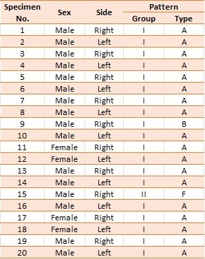
Table -2 shows the distribution of superficial palmar arch patterns observed in the current study. Out of the twenty hands, used in the current study, nineteen hands (95%) had complete superficial palmar arches while 1 hand (5%) had an incomplete arch. Out of the nineteen hands, eighteen (94.7%) were of type A (Classical radio-ulnar arch), while one hand (5.3%) was of type B (Ulnar type). The hand with an incomplete palmar arch exhibited Type A pattern (radio-ulnar type).
Table 2: Distribution of Superficial Palmar Arch patterns.
Vascular injection with RTV116:
The SPA patterns could be easily studied by vascular injection technique using RTV 116. This technique allows visualisation of minute branches which otherwise go unnoticed by the naked eye. RTV 116, being red in colour, allows a clear photographic record as well.
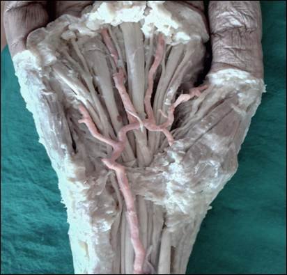
Fig. 3: Complete SPA formed by the union of radial and ulnar arteries.
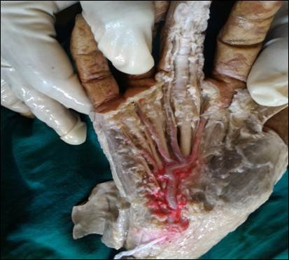
Fig. 4: Complete SPA formed by the ulnar artery only.
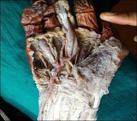
Fig. 5: Incomplete SPA formed by the radial and ulnar arteries.
DISCUSSION:
The human hand is gifted with a rich network of blood supply. Superficial palmar arch (SPA) and Deep Palmar Arch (DPA) contribute to the blood supply in the hand. The SPA and DPA are usually formed by two major arteries in the forearm, namely, radial artery (RA) and ulnar artery (UA), with their branches. Most of the times, the arteries which contribute to formation of SPA, form an anastomosis. Thus, the SPA may be classified as “complete” SPA. Sometimes, the anastomosis may be absent. In such cases, the SPA is termed as “incomplete” SPA [1].
Various authors have noted variations in the formation of the palmar arches and documented its prevalence, in literature. Proper identification and knowledge of such variations are essential especially during reconstructive or restorative hand surgeries so that a successful outcome can be ensured. Non-invasive methods such Doppler studies and angiography may be carried out to visualize the blood supply of the hand, but smaller caliber vessels may still go unnoticed. This is where, the need for an anatomical study of the arterial patterns in the hand, steps in.
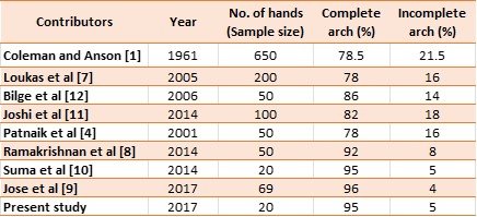
In the current study, 20 cadaveric hands were dissected and studied. Out of the 20 hands, a complete SPA, i.e arch formed by the anastomosis of contributing vessels, was found in 19 hands (95%). Only 1 hand (5%) had an incomplete SPA, i.e arch in which the contributing vessels do not anastomose. This finding is in accordance with most studies where a higher prevalence of complete SPA has been reported in comparison with incomplete SPA. Coleman and Anson [1] reported the prevalence of complete and incomplete SPA to be 78.5% and 21.5% respectively. Similarly, studies carried out by Jose et al [9], Ramakrishnan et al [8] , Suma et al [10], Joshi et al [11], Patnaik et al [4] , Loukas et al [7] and Bilge et al [12] noted a higher prevalence of complete SPA being 96%, 92%, 95%, 82%, 78%, 78% and 86% respectively.
On the contrary, a few authors like Sarkar et al [13], Fazan et al [14], Valeria et al [15] and Sullivan et al [16] have reported a higher incidence of incomplete palmar arch being 55%, 52%, 47.5% and 46.8% respectively.
Table 3: Comparison of SPA prevalence with previous studies.
The complete SPA can be classified, again, into five groups based on the arteries contributing to the formation of the arch.
In the current study, out of the 19 hands having complete SPA, 18 hands (94.7%) had a classical radio-ulnar arch, i.e. Type A, while 1 (5.3%) hand had ulnar dominant arch, i.e. Type B. The hand with incomplete SPA had non anastomosing radio-ulnar pattern, i.e. Type A. The findings of this study is similar to a study done by Madhyastha et al [17] who reported the incidence of Type A and Type B among the hands with a complete arch to be 93.75% and 2.08%. The incidence of Type F was 2.08%.
Table 4: Comparison of subtypes with previous studies.
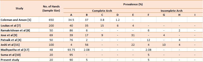
The most common type of complete arch found is the classical “radio-ulnar” pattern which is evident in the Table 4. However, Joshi et al [11] and Coleman & Anson [1] reported a higher percentage of complete arch to be of Type B, i.e. “ulnar” pattern. Among incomplete arch patterns, the “radio-ulnar” non-anastomosing pattern seems to be the most common.
It was also observed that there were different arches on the right and left hands of two cadavers. In one cadaver, both hands had a complete arch, but the right hand had Type B pattern and the left hand had Type A pattern. In another cadaver, the right hand exhibited pattern Type F (incomplete arch) and the left hand exhibited Type A (complete arch). Bilge and co-workers [12], in their study, also demonstrated a difference in arch patterns on the right and left sides in few cadavers.
Clinical implications:
The arterial arches anastomose freely with each other. As a result, any injury to the palmar or carpal arches may lead to severe bleeding with difficulty in maintaining haemostasis. It should also be noted that, apart from profuse bleeding, extensive vascular anastomosis in the hand can also bring about rapid wound healing [18].
There is a need to know the vascular patterns of the hand especially while making incisions to remove local pus from the hand. Accidental cutting of the superficial branch of the ulnar artery or the SPA can lead to profuse blood loss during the procedure.
Knowledge of the variations of SPA is not only helpful to the anatomists, but also to orthopaedic as well as microvascular surgeons during microvascular repair and re-implantation.
CONCLUSION
The human hand is far superior to that of the primates, as it is capable of performing complex movements. This can be attributed to the evolutionary modifications that have occurred over ages. Variations in vascular architecture of the hand have been discussed in literature and few authors have deduced that such variations could be remnants of primitive arterial patterns.
In the current study, out of the 20 cadaveric hands dissected, 19 hands (95%) presented with a complete superficial palmar arch (SPA) and 1 hand (5%) with incomplete SPA. Out of the 19 hands with complete arches, in 18 hands (94.7%), the SPA was formed by the radial and ulnar arteries. While in 1 hand (5.3%), the SPA was formed by ulnar artery (UA) alone. The hand with an incomplete SPA exhibited a radio-ulnar pattern.
It is not uncommon to find different SPA patterns in the same cadaver. Two cadavers, in the present study, showed this phenomenon. In one cadaver, the right hand had a complete SPA of Type B and the left hand had a complete SPA of Type A. In another cadaver, the right hand had an incomplete SPA of Type F and the left hand had a complete SPA of Type A.
The use of RTV 116 via vascular injection technique was beneficial to visualize the arch along with its minute branches and carry out meticulous dissection. Before injecting the flowable silicon, in this case, RTV 116, the body during embalming had to be injected with heparin solution so that the vessels were free from clots. Presence of clots can make the injecting of silicon into the vessel lumen very difficult.
The main limitation of the study was the sample size. Due to the limited availability of cadavers during the study period, only 10 cadavers could be utilized, providing 20 hands for the study. Hence, the frequency of variations observed was less.
AUTHOR CONTRIBUTIONS:
Pranup Roshan Quadras – Effective scientific and intellectual participation in the study; technical procedures; data acquisition; data interpretation; preparation and draft of the manuscript; critical review and final approval. Susie Jeyalyn David – Effective scientific and intellectual participation in the study; technical procedures; data interpretation; preparation and draft of the manuscript; critical review and final approval; statistical analysis. Prima Swetha Dsouza – Effective scientific and intellectual participation in the study; technical procedures; data acquisition; data interpretation; preparation and draft of the manuscript; critical review and final approval. Dane Chandy – Effective scientific and intellectual participation in the study; technical procedures; data acquisition; data interpretation; preparation and draft of the manuscript; critical review and final approval.
Conflicts of Interest: None
Cite this article: Pranup Roshan Quadras, Susie Jeyalyn David, Prima Swetha D’Souza, Dane Chandy. Mapping the Arterial Architecture of the Hand: A Cadaveric Study Using Vascular Silicone Gel Injection. Int J Anat Res. 2026;14(3):9646–9653. DOI: 10.16965/ijar.2026.184
References
- Coleman S, Anson B. Arterial patterns in the hand based upon a study of 650 specimens. Plastic and Reconstructive Surgery. 1962;29(1):85-86. https://doi.org/10.1097/00006534-196201000-00028
- Gellman H, Botte M, Shankwiler J, Gelberman R. Arterial Patterns of the Deep and Superficial Palmar Arches. Clinical Orthopaedics and Related Research. 2001;383:41-46. https://doi.org/10.1097/00003086-200102000-00007 PMid:11210968
- Moore K, Dalley A, Agur A. Upper Limb. Clinically Oriented Anatomy. 13th ed. Lippincott Williams & Wilkins; 2013. p. 781-782.
- Patnaik, V.V.G, Kalsey G, Singla R K. Palmar arterial arches-A morphological study. Journal of Anatomical Society India.2001;51(2):187-193.
- Ottone N E, Prum N, Dominguez M, Medan C et al. Analysis and clinical importance of superficial arterial palmar irrigation and its variation over 86 cases.International Journal of Morphology. 2010; 28(1):157-164. https://doi.org/10.4067/S0717-95022010000100022
- Rodríguez-Niedenführ M, Burton GJ, Deu J, Sañudo JR. Development of the arterial pattern in the upper limb of staged human embryos: normal development and anatomic variations. J Anat. 2001 Oct; 199 (Pt. 4):407-17. https://doi.org/10.1046/j.1469-7580.2001.19940407.x PMid:11693301 PMCid:PMC1468351
- Loukas M, Holdman D, Holdman S. Anatomical variations of the superficial and deep palmar arches. Folia Morphol (Warsz). 2005 May;64(2):78-83.
- Vidya R, Anil kumar R, Aruna S et al. A cadaveric study on anatomical variations of the superficial palmar arch. Int. J Med Res Health Sci.2014;3(1):144-148. https://doi.org/10.5958/j.2319-5886.3.1.028
- Jose BA, Shashi Rekha M, Babu S. Analysis of the vascular pattern in the superficial palmar arch formation. Indian Journal of Clinical Anatomy and Physiology. 2017;4(1):11-5.
- Suma MP, Kumar VS, Ranganath P. An Anatomical Study of Superficial Palmar Arch. Int J Anat Res. 2014; 2(4): 725-39. https://doi.org/10.16965/ijar.2014.536
- Joshi SB, Vatsalaswamy P, Bahetee BH. Variation in formation of superficial palmar arches with clinical implications. Journal of clinical and diagnostic research: JCDR. 2014 Apr;8(4):AC06. https://doi.org/10.7860/JCDR/2014/7078.4252 PMid:24959427
- Bilge O, Pinar Y, Ozer MA, Gövsa F. A morphometric study on the superficial palmar arch of the hand. Surg Radiol Anat. 2006 Aug;28(4):343-50. https://doi.org/10.1007/s00276-006-0109-9 PMid:16642281
- Sarkar A, Dutta S, Bal K, Biswas J. Handedness may be related to variations in palmar arterial arches in humans. Singapore Med J. 2012 Jun 1;53(6):409-12.
- Fazan V, Borges C, Silva J et al. Superficial palmar arch: an arterial diameter study. J. Anat.2004;204:307-11. https://doi.org/10.1111/j.0021-8782.2004.00281.x PMid:15061757 PMCid:PMC1571293
- Valeria P S F, Celso T B, Jefferson H S, Abadio G C, Omar A R F. Superficial palmar arch: an arterial diameter study. J. Anat. 2004; 204: 307 -311. https://doi.org/10.1111/j.0021-8782.2004.00281.x PMid:15061757 PMCid:PMC1571293
- Elizabeth O' Sullivan, Barry S Mitchell. Association of the absence of Palmaris longus tendon with an anomalous superficial palmar arch in the human hand. Journal of Anatomy.2002;202 (2): 253 -257.
- Madhyastha S, Murlimanju BV, Jiji PJ, Saralaya VV, Rai A, Vadgaonkar R. Morphological variants of the human superficial palmar arch and their clinical implications. Journal of Morphological Science. 2011;28(4):261-4.
- Lockhardt RD, Hamilton GF, Fyfe FW. Anatomy of the human Body. London : Feber & Feber Ltd; 1959, p. 612-619.


