
Exploring the Variations of the Cavernous Sinus Tributaries in the South African Population
Thembelihle Cele, Sodiq Kolawole Lawal , Samuel Oluwaseun Olojede , Okikioluwa Stephen Aladeyelu, Carmen Olivia Rennie · Int J Anat Res 2026;14(3):9584–9598
1 Discipline of Clinical Anatomy, College of Health Sciences, Nelson R. Mandela School of Medicine campus, University of KwaZulu-Natal, Durban, KwaZulu-Natal, 4000, South Africa.
2 Department of Anatomy and Physiology, School of Biomedical Science and Pharmacy, University of Botswana, Private Bag 0022, Plot 4775, Notswane Road, Gaborone, Botswana.
3 Division of Human Anatomy, School of Biomedical Sciences, Faculty of Medicine and Health Sciences, Walter Sisulu University, Mthatha, 5099, South Africa.
4 Discipline of Clinical Anatomy, College of Health Sciences, Westville campus, University of KwaZulu-Natal, Durban, KwaZulu-Natal, 4000, South Africa.
5 Department of Natural and Rehabilitative Sciences, Faculty of Health Sciences, University of Fort Hare, East London, South Africa.
Corresponding Author: Thembelihle Cele, Discipline of Clinical Anatomy, School of Laboratory Medicine and Medical Sciences, College of Health Sciences, Nelson Mandela School of Medicine, University of KwaZulu-Natal, Durban, KwaZulu-Natal, 4000, South Africa. · Thembeh2019@gmail.com
Abstract
Background
The cavernous sinus (CS), being the site of thrombosis, predisposes most patients to life-threatening diseases of the cavernous sinus and cerebral venous system. The procedure to treat these life-threatening diseases requires a good anatomical knowledge of CS tributaries owing to their variations in morphological and drainage patterns.
Methods
One hundred CT images of males and females were used (≥13 years). In this study, the sample drawn from the Black South African population was compared to the sample drawn from the Indian and White SA population groups. The full length of each vein - the superior and inferior ophthalmic veins (SOV and IOV), the sphenoparietal sinus (SPS), and the superficial middle cerebral vein (SMCV), were observed and classified into various types according to their drainage into the cavernous sinus. These types were compared to factors such as population, age, sex and laterality.
Results
Three morphological variations were observed for the SOV, Type S was the most prevalent in the Indian/White sample group on both sides, and more common in females. For the IOV, Type A was the most prevalent in all parameters studied. Regarding the SPS, Type A was the most prevalent, followed by Type B, and Type C was the lowest recorded. For the SMCV, Type C was the most prevalent, followed by Types A and B, with less frequencies recorded for Type D. A statistically significant association for the drainage pattern of the IOV was found on the right side for sex (p = 0.03) only.
Conclusion
The SOV showed less variations and may be an ideal vein to be used for transvenous embolisation. The use of CT images to study the pattern and variations of these veins is highly recommended before transvenous embolisation.
Background
The cavernous sinus (CS) is known as a frequent site of thrombosis, which is a process of formation of blood clots in vessels in response to an infection from the face or the orbit called cellulitis [1; 2]. Thrombosis of the CS can result in inflammation of the adjacent neurovascular structures, while diseases such as cavernous sinus thrombosis (CST), which are life-threatening, can result in the formation of blood clots in the CS [2]. The aetiology of these diseases could be as a result of an infection from the face, sinuses, orbital cellulitis, and pharyngitis, but they are predominantly found in patients with diabetes and thrombophilia [2]. These conditions are reported to predominantly affect women of reproductive age [2]. CST can cause long-term damage to the brain, eyes, and nerves, and without sufficient treatment, may lead to death [2].
The CS is also a site of dural arteriovenous fistulae (dAVF), an abnormal connection between the internal or external carotid arteries, which results in the CS being fed by arteries [3]. This type of malformation is often as a result of traumatic brain injury, embryological defects, surgery, and/or genetic conditions [3]. This condition is normally found unilaterally; however, the majority of females were found to possess bilaterality (90%, p = 0.043) of this condition. Although it is also found unilaterally in males (10%) [3].
Among the life-threatening disorders affecting the CS is cerebral venous sinus thrombosis (CVST), a type of stroke normally found in women in their reproductive ages [4]. Although the exact aetiology of the disease is not known, it is found to affect women on oral contraceptives, during pregnancy, and in the peripartum period [5; 6]. Previous literature has documented the use of contraceptives in relation to CVST, and a significant association was obtained, which confirmed that females are the most vulnerable to this disease [4]. A systematic review study on the CVST in sub-Saharan Africa found a very high prevalence of women with CVST (58.6%) [6]. In a case series report of 210 patients, hemicraniectomy was successfully done in one patient (2.1%); meanwhile, ten percent of the patients died after the surgery, with 46.5% of the patients recovering either completely or partially [6].
Surgical interventions to these cerebral venous sinus diseases in the skull base have resulted in very high mortality and morbidity rates (10%) in sub-Saharan Africa [5,6]. This is owing to the intracranial bleeding caused by the rupture of veins during these surgical procedures [5-7].
Endovascular embolisation has been the treatment of choice for this condition because it is safer and possesses low complication rates [3]. The access to the CS when performing the endovascular embolisation is through the inferior petrosal sinus (IPS), but if it is occluded, the point of access will be through the facial and ophthalmic veins [3]. Although the endovascular embolisation procedure is declared safe and presents fewer complications, it is important to note that the veins used in accessing the CS have numerous variations in their morphology and drainage patterns to the CS, thus making the procedure difficult [8].
The CS receives communication from numerous para-cavernous sinus structures such as the superior and inferior ophthalmic veins [1; 9; 10]. The superior ophthalmic vein (SOV) runs between the optic nerve and the superior rectus muscle and exits the orbit via the superior orbital fissure to drain into the CS [11,12]. However, Tsutsumi et al. [12] who observed the course of the SOV from distal to proximal using magnetic resonance angiography (MRA) found that morphological variations in terms of laterality were evident. The proximal segment of the SOV was found to be tortuous in seven percent of the population on the left side and in eleven percent of the population on the right [12].
Furthermore, the inferior ophthalmic vein (IOV), which is located in the midorbit slightly superior to the inferior rectus muscle, was found to drain into the CS in two different patterns [11]. The IOV either curved upwards laterally and infrequently medial to the optic nerve to join the third part of the SOV and then drained into the CS, or it drained directly into the CS, bypassing the SOV [11]. These observations were not compared according to sex, age and laterality.
The sphenoparietal sinus (SPS) is known to receive blood from the superficial middle cerebral vein (SMCV) located in the lesser wing of sphenoid bone before it emptied into the CS [9]. They found the SPS to drain in three different patterns using Magnetic Resonance (MR) images and the patterns were classified into types, namely: Type A- drained into the anterior aspect of the CS; Type B- connected with the sphenoid emissary veins (SEV) or the pterygoid plexus; and Type C- the hypoplastic (underdeveloped) SPS drained into the anterior aspect of the CS.
As mentioned above, the SMCV empties into the SPS [9]. However, different studies have noted variations in the drainage pattern of the SMCV [9; 13]. Suzuki and Matsumoto [13] observed seven different drainage patterns of the SMCV. Meanwhile, previous findings by Tanoue et al. [9] in their study observed only four types: Type A- the SMCV drained into the SPS, Type B- entered the anterior aspect of the CS, Type C- connected to the SEVs to drain into the pterygoid plexus and Type D- emptied into the superior petrosal sinus/transverse sinus. Type A was the most prevalent, followed by TypeB. Two percent of the subjects presented with mixed types [9].
Although, there is existing literature regarding the variations of the CS tributaries, it is important to note that variations regarding the age, sex, laterality in the SA population remain undocumented. Also, the use of CT images to study the pattern and variations of these veins is highly recommended before transvenous embolisation is performed. Therefore, the current study, using CT images aimed to report on the variable patterns of the CS tributaries in the SA population with comparison to sex, age, and laterality.
Materials and Methods
Sample size: This was a retrospective analysis of 100 CT scans of adult individuals (56 males, 44 females). The left and right sides were assessed separately, amounting to a total of 200 sides. The CT images were obtained from a tertiary hospital, the Inkosi Albert Luthuli Central Hospital (IALCH) in Durban, Kwa-Zulu Natal, South Africa.
Age classification and population: The CT images were of 13 years of age and above. The age categories were classified according to Biwasaka et al. [14] as follows: <29, 30-39, 40-49, 50-59, 60-69, 70-79, 80-89, and >89 years of age. The starting age was considered because the venous sinuses of the skull demonstrate their most significant growth between the ages of zero and seven years and typically reach adult size around five to ten years of age [15]. The diseases affecting the dural venous sinuses were also found to affect women in their reproductive ages (age ≥ 13 years) [4].
Regarding population, due to the skewness of data in the CT sample, the White and the Indian sample groups were merged into one sample and were compared to the Black sample, ‘Black SA vs. Other SA sample groups (Indian/White SA)’.
The composition of the sample for the CT images were Black (71; 71%) and Indian/White (29; 29%). (Note: Generally, the distribution of the SA population, Black SA makes up about 79.8%; White SA makes up about 8.7%, with Indian SA making up about 11.5%) [16,17].
Materials
A total of 100 CT images from CT venograms and CT angiograms (with venous phase) were used in the study. The digital imaging and communications in medicine (DICOM) images were viewed and analyzed using syngo.plaza viewer software (version VB10). The morphology and drainage patterns of the veins were viewed in axial and sagittal planes.
Scanning protocol
The CT images were acquired using Multi-Detector row CT(MDCT) Scanners (Lightspeed CT, GE Healthcare Medical System, Milwaukee, Wisconsin, USA, and SOMATOM Definition Flash CT Scanner, Siemens Healthineers, Forcheim, Germany, both of 1mm and 2 mm slice configuration, respectively). Acquisition number: 18; CS:.60; Exposure Time:285; KVP:120.00; X-ray tube Current:425; Feed: 46.00.
Inclusion and exclusion criteria
The inclusion criteria for scans were as follows:
- CT images of patients who were ≥ 13 years of age, depicting both cerebral hemispheres.
- CT images with a slice thickness of either 1 or 2 mm.
The exclusion criteria for scans were as follows:
- Scans with observable pathologies affecting the venous system, e.g., the arteriovenous malformation, dural arteriovenous fistula, brain tumours, and veno-occlusive diseases, were excluded from the study.
Ethical clearance
Ethical clearance was obtained from the Biomedical Research Ethics Committee (BREC) at the University of KwaZulu-Natal (BREC/00005683/2023). Permission to access CT scans and conduct this research was sought and approved by the institution (UKZN) and the Department of Health, KwaZulu-Natal.
Study procedure: The DICOM images were viewed on the syngo.plaza viewer software (version VB10). The morphology and drainage patterns of the CS tributaries were viewed in two views/planes (axial and sagittal), and each tributary was analyzed as follows:
Superior ophthalmic vein (SOV): The morphology (tortuosity) of the SOV was observed from its confluence with the supraorbital and angular veins to its entrance in the CS. To observe the course of the SOV, axial CT images were used. To identify the SOV, the location of the frontal bone (FB) and frontal sinus (FS) were used as landmarks in identifying the SOV because the FS is drained by the SOV, which enters the orbit through the superior orbital foramen (SOF) [18]. They were also found to course laterally to the lamina papyracea under the superior rectus muscle [18]. These two landmarks (FB and FS) were used, and the SOV was clearly observable at this level. To identify tortuosity, a tortuous pattern was assessed on the anterior and posterior ends of the SOV.
Inferior ophthalmic vein (IOV): The drainage pattern of the IOV was observed from the anterior part of the floor of the orbit to its entrance in the SOV or directly into the CS. To identify the IOV, sagittal CT images were used. The floor of the orbit and the optic nerve were used as landmarks. This is because the IOV travels at the floor of the orbit inferior to the optic nerve until it enters the middle cranial fossa through the superior orbital fissure just inferior to the common tendinous ring [11].
Sphenoparietal sinus (SPS): The SPS was observed from the junction of the frontal and temporal bone (pterion) to its entrance in the CS or at a point where it converges with the SEVs. To observe the presence of the SPS, axial CT images were used, and the sinus usually appears as a curved and contrast-enhancing structure. The posterior edge of the lesser wing of the sphenoid bone was used as a landmark as the SPS travels inferior to this structure to enter the CS [9].
Superficial Middle Cerebral Vein (SMCV): The drainage pattern of the SMCV was observed from the temporal region of each hemisphere to its drainage in the CS, SPS, and the superior petrosal/transverse sinus, as well as the SEVs. To identify the SMCV, axial CT images were used. To trace the path of the SMCV, the insula was used as the landmark since the SMCV courses on the lateral sulcus (Sylvian fissure), which is found lateral to the insula. It moves in a posterior-anterior direction and curves anteriorly at the tip of the temporal lobe to drain into the SPS or directly into the CS [13]. Hence, the insula was used as a landmark.
Statistical analysis:
The Stata version 17 (StataCorp, College Station, Texas, USA) statistical software was used for analysis. Statistically significant level was set at p<0.05. Descriptive statistics were used to summarise the data. Frequencies and percentages were used for categorical data. Chi-square tests were used to identify demographic characteristics (population, sex and age) associated with the CS. The Mann-Whitney (2 variables) and Kruskal-Wallis (more than 2 variables) non-parametric test were used.
Results
The superior ophthalmic vein (SOV)
This study assessed the morphology of the SOV from the confluence of the supraorbital and angular veins to its entrance in the CS. Three morphological patterns of the SOV were observed: (a) Type S: the SOV was straight from anterior to posterior, (b) Type T-A: the SOV was tortuous anteriorly just posterior to the eyeball, and (c) Type T-P: the SOV was tortuous posteriorly, towards its entrance in the CS. The left and right sides were assessed separately.
Table 1: Showing CTs with the same configuration of the superior ophthalmic vein on the left and right sides (%).
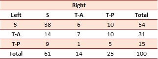
Out of the 100 individuals assessed in the study, some individuals showed the same pattern on the left and right side. Thirty-eight percent of CTs accounted for Type S, seven percent accounted for Type T-A and five percent accounted for Type T-P. These percentages amounted to 50% of the population, meaning that 50% of the population had a mixture of either Type S, T-A and T-P.
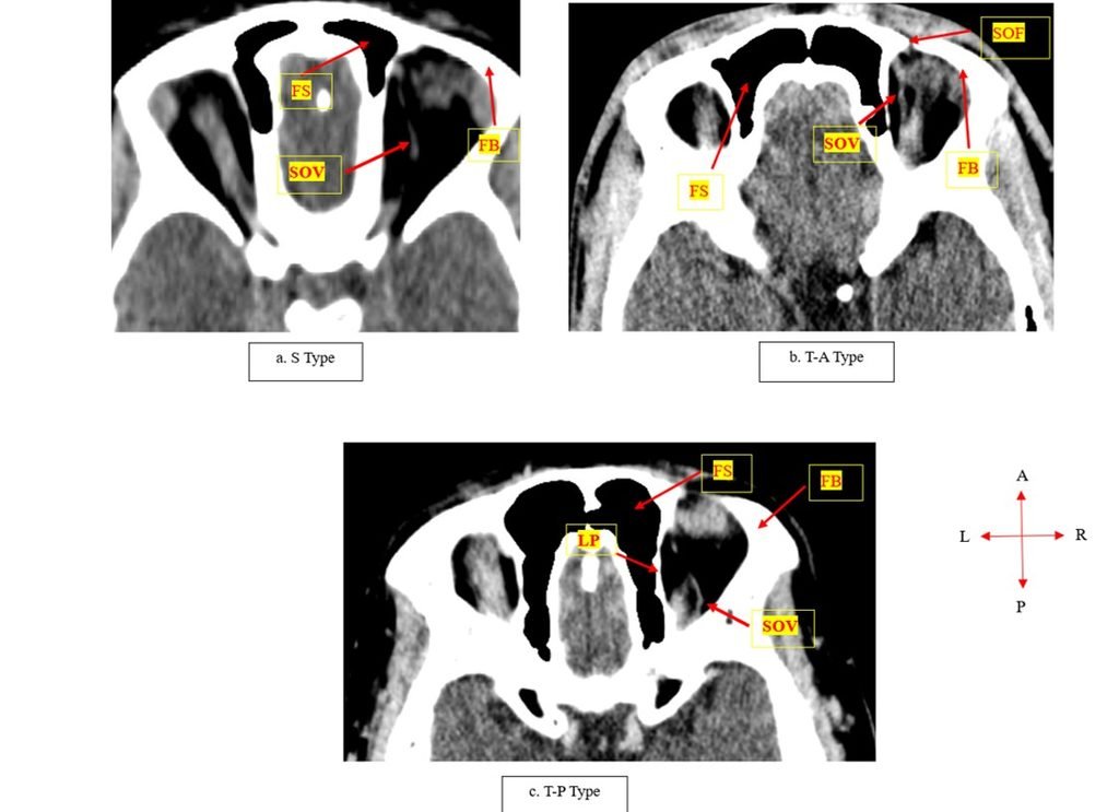
Fig. 1: Axial CT images showing the different morphological patterns of the superior ophthalmic vein; S Type – straight from proximal to distal; Type T-A – tortuous anteriorly (distally); and Type T-P – tortuous posteriorly (proximally) (images by Cele et al., 2026).
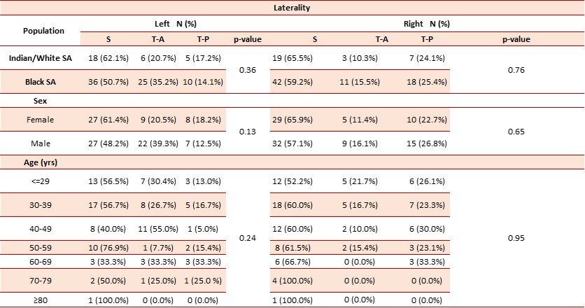
Table 2: The morphology of the SOV compared to population, sex, age, and laterality.
The prevalence of SOV and Population
The Black sample group was compared to the Indian and White sample groups (for the purposes of convenience, the Indian/White sample was combined). The sides (left and right) were assessed separately. Type S was the most prevalent in both sample groups on both sides with the Black sample accounting for 50.7% on the left side and 59.2% on the right side, whistle the Indian/White sample accounted for 62.1% on the left and 65.5% on the right side. Type T-A was the second most prevalent on the left side and Type T-P on the right side for both sample groups. There was no statistical significance found on either side as indicated in Table 2.
Prevalence of SOV and Sex
Type S was the most prevalent on both the right and the left sides and in both sexes. Females presented with a high incidence of Type S compared to males on both sides. Type T-A was the second most prevalent on the left side, and Type T-P on the right side for both males and females. However, males presented with high incidence of Type T-A on the left side and Type T-P on the right side compared to females. Overall, there was no statistical significance association between the SOV types and sex.
Prevalence of SOV and Age
The age groups were divided into seven groups with a 10-year age gap. A trend was noted amongst the three types and the age groups, where the prevalence was high in the first three age groups; the prevalence of each type decreased with increasing age on both the left and the right sides, it increased again with age advancement. No statistical significance was found for age as indicated by Table 2.
The inferior ophthalmic vein (IOV)
The study looked at the drainage pattern of the IOV. Two drainage patterns were found:Type A- the IOV drained into the SOV before draining into the CS, Type B- the IOV drained directly to the CS, bypassing the SOV.
Table 3: Showing patients with the same pattern of the inferior ophthalmic vein on the left and right sides (%).
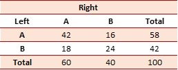
Out of the 100 individuals assessed in the study, 42% of individuals presented with Type A on both sides, while 24% presented with Type B on both sides. This makes 66% of the population, and the remaining 34% of the population had a mixture of both Type A and B on the left and right sides.
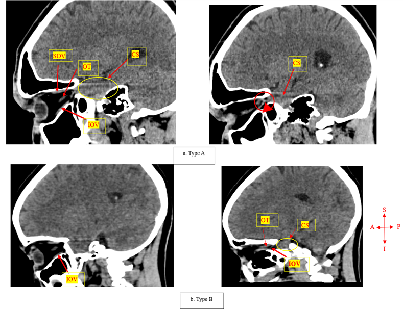
Fig. 2: Sagittal CT images showing the different drainage patterns of the inferior ophthalmic vein; Type A – drains into the superior ophthalmic vein before draining into the cavernous sinus; Type B – drained directly into the cavernous sinus bypassing the superior ophthalmic vein (images by Cele et al., 2026).
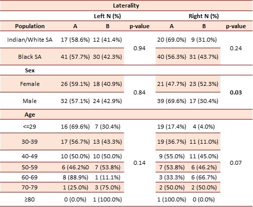
Table 4: The drainage pattern of the IOV compared to population, sex, age, and laterality.
The drainage pattern of the IOV and Population
The Black sample group presented with high incidence of Type B, while the Indian/White sample group presented with high incidence of Type A on the left and right sides. There was no statistical significance found between population and sides. Overall, Type A was the most prevalent on both sides in both sample groups compared to Type B as illustrated in Table 4.
The drainage pattern of IOV and Sex
Type A had a higher incidence on the left side for both sexes, with males having more Type B and less Type A compared to females. Similarly on the right side, females presented with a high prevalence of Type B and less of Type A. There was a statistically significant association found on the left side regarding sex, p = 0.03.
The drainage pattern of IOV and Age
A high prevalence of Type A was noted in the ≤29 years and 30-39 years age groups on the left side compared to the right side. Fifty percent of Types A and B were found in the 40-49 years age group on the left side, and in the 70-79 years age group on the right side. Only 1 CT was found in the ≥80 years age group, and the CT accounted for 100% of Type A on the right and Type B on the left side as shown in Table 4.
Sphenoparietal sinus (SPS)
The drainage pattern of the SPS was assessed and three types were observed: Type A- the long SPS drained into CS only; Type B- the SPS drained into the CS and the pterygoid plexus, and Type C- the SPS was hypoplastic (poorly formed).
Table 5: CTs with the same configuration of the sphenoparietal sinus on the left and right sides (%)
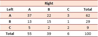
A total of 100 individuals were assessed in the study. Regarding CTs that presented the same veins on both sides; 37% accounted for Type A, 15% individuals accounted for Type B, and two percent accounted for Type C. This makes up 54% of the total population; the rest of the CT had a mixture of either Type A, B, or C.
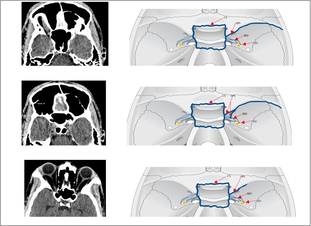
Fig. 3: Axial CT images illustrating the different drainage patterns of the sphenoparietal sinus; Type A – drained into the lateral aspect of the cavernous sinus; Type B – it connected to the cavernous sinus and the sphenoid emissary veins; Type C – is hypoplastic (images by Cele et al., 2026).
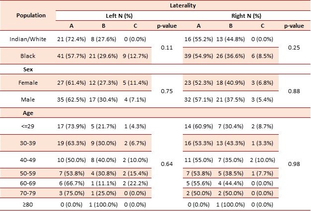
Table 6: The drainage pattern of the sphenoparietal sinus compared to population, sex, age, and laterality.
The drainage pattern of the sphenoparietal sinus and Population
A high prevalence of Type A was found on the left side for the Black and Indian/White sample groups. The Indian/White sample had a high prevalence of Type A and B, and none of the CTs presented with Type C in this sample group on the right side. The Black sample showed consistency in terms of presence in each type. No statistical significance was found on the left and right sides about population p = 0.11 and 0.25, respectively, as shown in Table 6.
The drainage pattern of the sphenoparietal sinus and Sex
Type A was the most prevalent on the left and right sides in both sexes. Type B was the second most prevalent in both sexes and on both sides, with Type C being the lowest prevalence. Males had a higher prevalence of Type A compared to females. There was no statistically significant association found on the left and right sides regarding sex.
The drainage pattern of the sphenoparietal sinus and Age
High prevalences of Types A and B were noted from the ≤29 years to 40-49 years age groups; however, from the 50-59 years age group, a decrease in prevalence on both sides was observed. Furthermore, one CT was found in the ≥80 years age group, and the CT accounted for 100% of Type B on both sides as shown in Table 6. There was no statistically significant association found for age.
Superficial middle cerebral vein (SMCV)
The drainage pattern of the SMCV was examined, and four patterns were observed: Type A- the SMCV drained into the SPS before draining into the CS; Type B- the SMCV drained laterally into the CS; Type C- the SMCV connected to the SEV and drained into the pterygoid plexus of veins; and Type D- the SMCV drained into the superior petrosal/transverse sinus posteriorly. These types are indicated in the Table below:
Table 7: Showing CT with the same pattern of the superficial middle cerebral vein (%).
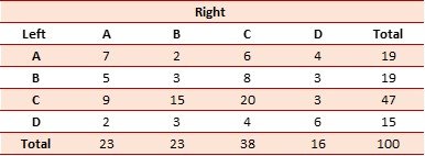
Out of 100 CT images, seven percent presented with Type A on both sides, three percent presented with Type B, 20% presented with Type C, and six percent presented with Type D. This makes 36% of the total population, the rest of the population had a mixture of either Types A, B, C or D.
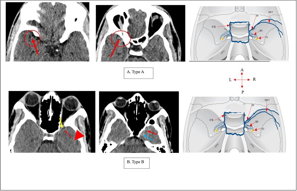
Fig. 4: Axial CT images illustrating the different drainage patterns of the superficial middle cerebral vein: Type A – the superficial middle cerebral vein drained into the sphenoparietal sinus; Type B – it drained into the lateral aspect of the cavernous sinus; Type C – it connected to the sphenoid emissary veins draining into the pterygoid plexus of veins; and Type D – it drained into the superior petrosal sinus and the transverse/sigmoid sinus (images by Cele et al., 2026).
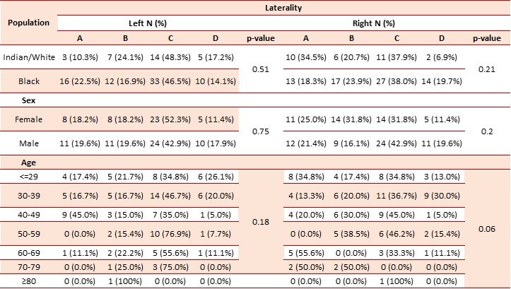
Table 8: The drainage pattern of the superficial middle cerebral vein compared to population, sex, age, and laterality.
The drainage pattern of the superficial middle cerebral vein and Population
Type C was the most prevalent on both the left and the right sides in both sample groups, with Type D being the lowest recorded. Type A was the second most prevalent on the right side and Type B on the left side as shown in Table 8. There was no statistical significance found on the right or the left side regarding population.
The drainage pattern of the superficial middle cerebral vein and Sex
A high prevalence of Type C was found on both sexes and on both sides. Equal numbers for Types A and B were found for males (19.6%) and females (18.2%) on the left side. Females presented with equal numbers of Types B and C (n= 31.8%) on the right side. Furthermore, females presented with the same incidence for Type D on both sides (11.4%), with males also presenting with the same percentage of Type C on both sides (42.5%) as shown in Table 8. No statistically significant association was found on both sides for sex.
The drainage pattern of the superficial middle cerebral vein and Age
Among all the types, Type C was the most prevalent on the left and right sides. Type B showed consistency in terms of prevalence across all age groups with Type D having the lowest frequencies recorded. The 70-79 years age group presented with 50% of Types A and B. No statistical significance was found on the left and right-side regarding age.
Discussion
Diseases of the dural venous sinuses, such as the CST and CVST, are life-threatening disorders that result in the formation of blood clots in the CS [3,4,19]. These diseases have been linked to high morbidity and mortality rates [5]. Additionally, the CS is also a site of dAVF that are said to be treated with endovascular embolisation [20]. This is the safest and most effective method for managing CS diseases that require surgical intervention [20]. The transvenous type of embolisation uses the IPS to access the CS [21]. However, if the IPS is obstructed, the SOV may be used as an alternative [21,22].
The superior ophthalmic vein
Several studies have observed the tortuosity of the SOV, which sometimes becomes an obstruction in the access of the CS using the transvenous approach [12,14,22-24]. The study by Kurata et al. [22] observed the tortuosity of the SOV in three of the ten patients who were examined. The tortuosity of the SOV was observed distally on the left side in those three patients [22]. This is in line with the current study, as the left side had a high incidence of SOV with Type T-A (tortuous anteriorly) compared to the right side in sex, population, and age. Both sides can have different morphological patterns of the SOV; therefore, assessment of the SOV is crucial before performing transvenous embolization.
In a study by Wolfe et al. [23], out of the ten patients that were studied, one CT presented with tortuosity of the SOV on the left side, which resulted in the rupture of the vein during the direct surgical SOV approach as well as the retrograde transvenous embolization procedure. Despite this incident, they still recommend the use of the SOV as an alternative in accessing the CS by direct surgical cannulation if the IPS, superior petrosal sinus, and the intercavernous sinus are obstructed [23]. This agrees with the present study as the tortuosity of the SOV both anteriorly and posteriorly was less compared to the straight pattern of the SOV. This implies that the SOV can be used for transvenous embolisation in the SA population as this will limit the amount of venous rupture and intracranial bleeding during surgery.
Additionally, the study by Tsutsumi et al. [12] observed the whole course of the SOV, and its proximal part was found to be variable. Out of 46 patients examined, five (11%) patients had a tortuous proximal segment on the right side, and three (6.5%) patients presented with a tortuous proximal segment on the left side. This also agrees with the present study, as variations between the left and right sides were evident. This suggests that each CT may present with different patterns of the SOV on the left and right sides.
The inferior ophthalmic vein
Venous sinuses are often assumed to facilitate the spread of infection from extracranial to intracranial by the SOV and IOV to the CS, which in turn leads to life-threatening septic cavernous thrombosis [26-28]. The IOV is said to converge with the SOV before draining into the CS [11,26,29]. Zhang and Stringer [26] observed a similar drainage pattern of the IOV in their study; however, the prevalence of these findings was not reported. Cheung and McNab [11] also observed a similar pattern in three out of ten orbital dissections.
This is in agreement with the current study as the prevalence of Type A was high when compared to Type B in both population groups and both sexes, with a statistical significance of p = 0.03 found on the right for sex. It is worth noting that Cheung and McNab (2003) observed the IOV draining directly into the CS, bypassing the SOV in two orbital dissections. Similar to the current study, low incidents of Type B were obtained for population, sex, and age. This implies that most individuals in SA are likely to present with Type A, and this can result in complications like embolisation of unintended areas as well as damage to surrounding structures like rupture of the SOV.
The sphenoparietal sinus
The SPS has been identified as a venous channel found under the lesser wing of the sphenoid bone, and it receives drainage from the SMCV [30; 31]. The relationship between the SPS and the SMCV in terms of their drainage has been studied previously [9,13,30]. However, these studies were more concerned about the drainage of the SMCV and less on the SPS.
Tanoue et al. [9] observed three different patterns of the SPS which were also observed in the current study. Out of the 37 patients that were assessed, Type A (72%) was the most prevalent, followed by Type C (14%), and Type B (4%) was the least prevalent in the Japanese study [9]. The current study agrees with the Tanoue et al., [9] as Type A was the most prevalent in all the parameters (population, sex, and age) studied on both the left and right sides. However, Type A was followed by Type B, and fewer incidences were recorded in the present study for Type C. It is important to note that the Indian/White sample group did not present with Type C on both the left and right sides. This assumes that it is possible to access the CS using the SPS in the Indian/White sample group, as the course of the SPS was observed in most patients coursing from the pterion along the lesser wing of the sphenoid to its termination in the CS (Type A).
The superficial middle cerebral vein
The SMCV is one of the venous channels essential in the transvenous embolisation of the cavernous sinus dural arteriovenous fistula (CSDAF) [31]. It has been observed in previous studies, and it was said to drain either to the SPS before draining into the CS or just directly to the CS (9; 13; 31]. However, some authors have noted several drainage patterns of the SMCV to the CS. A study of 24 patients observed four drainage patterns where the SMCV drained into the anterolateral aspect of the CS in 35.4% of the patients; drained into the foramen ovale in 10.4% of the patients; 16.7% of the population had their SMCV draining into the lateral aspect of the CS and in 35.4% of the population they could not identify the SMCV [31]. Suzuki and Matsumoto [13] and Ikushima et al. [33] observed seven drainage patterns of the SMCV, which were classified according to their termination points as SPS, CS, Superior petrosal sinus, basal type, squamosal type, as well as the underdeveloped type.
The studies by Suzuki and Matsumoto [13] and Ikushima et al. [33] observed a high prevalence of the SPS type in their studies – 58% and 54% respectively. Tanoue et al. [9] observed four drainage patterns of the SMCV as observed by the present study and were classified into Type A – SPS, Type B – CS, Type C – SEV, and Type D – Superior petrosal sinus. Type A was the most prevalent in their study with Type D being the least recorded [9]. Contrary to the present study, Type C was the most prevalent, but Type D was the least recorded similar to previous studies, with notable differences on the left and right sides.
There were similar findings for Types A and B for females and males on the left side (18.2% and 19.6%, respectively). Similarly, in previous studies, patients with the same or mixed types on the left and right sides were observed as indicated in Table 7 [9]. This implies that the pattern of the SMCV may be challenging to use for transvenous access to the CS as it is small and variable. It can pose a high risk to surrounding structures like the middle meningeal artery, but it can be used as a potential access point for malformations and CSDAF. However, this will depend on individual patients’ anatomy as the SMCV may not drain directly to the CS but drain to the pterygoid plexus through the SEVs and to the superior petrosal sinus/transverse sinus.
Knowledge of the drainage patterns of the tributaries draining into the CS is essential in the transvenous embolisation of diseases affecting the CS. The SOV may be used for transvenous access to the CS, as it presented with less variations in terms of morphology. However, if not assessed properly, it may result into serious clinical complications. Therefore, adequate CT analysis must be done before attempting to access the CS using the SOV. The IOV may be used depending on an individual’s venous anatomy. If it has a high incidence of Type A, it could cause embolisation of the SOV and neighbouring structure if it is used to access the CS. To prevent cortical venous reflux into the CS during transvenous embolisation, it is important for clinicians to be aware of the drainage patterns of the SMCV as well as its connection with SPS.
Variations of the CS tributaries were noted across different population groups, sex, as well as age. Therefore, the use of CT images in the assessment of cerebral venous anatomy and diagnosis of venous sinus diseases is essential in clinical settings before deciding on the appropriate treatment method. Although there was a slight difference with regards to sex across all the veins studied, it was statistically significant: IOV (p = 0.03). These variations must be considered in the diagnosis and treatment of venous sinus diseases. One CT in the ≥ 80 years age group was found to have variations on the left and right sides. However, this CT presented with same pattern for the SOV (S Type) and SPS (Type B) on the left and right sides. However, for the IOV and the SMCV, the same CT presented with different patterns on the left and right sides: IOV (left – Type B: right – Type A) and SMCV (left – Type B: right – Type C). This shows that different veins may be used to access the CS for the same patient, as there may present with variations of the same vein on both sides.
Conclusion
In this study population, the IOV, SPS, and SMCVexhibited significant anatomical variability, posing a risk of clinical complications during transvenous embolisation. Unless a patient’s specific anatomy is uniquely favourable, these vessels are generally unsuitable targets. Conversely, the SOV demonstrated greater consistency and remains a more viable option. Regardless of the target vessel, the use of pre-procedural CT imaging is highly recommended to map individual venous patterns and mitigate risks.
Limitations
This study was limited by population diversity owing to the use of a single public hospital. Future studies can be done on the course and drainage patterns of the CS tributaries by collecting from different hospitals to allow for population diversity.
List of Abbreviations
CS – cavernous sinus
CST – cavernous sinus thrombosis
dAVF- dural arteriovenous fistulae
CVST – cerebral venous sinus thrombosis (CVST)
IPS – inferior petrosal sinus
MRA – magnetic resonance angiography
SOV – superior ophthalmic vein
IOV – inferior ophthalmic vein
SPS – sphenoparietal sinus
SMCV – superficial middle cerebral vein
SEV – sphenoid emissary veins
IALCH – Inkosi Albert Luthuli Central Hospital
SA – South Africa
CT – computed tomography
FB – frontal bone
FS – frontal sinus
SOF – superior orbital foramen
CSDAF – cavernous sinus dural arteriovenous fistula
Conflicts of interest
Conflicts of interest: The authors declare that they have no conflicts of interest.
Author contribution
Thembelihle Cele, Okikioluwa Stephen Aladeyelu, Sodiq Kolawole Lawal, Samuel Oluwaseun Olojede, Carmen Olivia Rennie. T and CO designed the experiments, T performed the experiments and collected data, T, CO, OS, SK, and SO discussed the results and strategy, CO supervised, directed, and managed the study, CO, OS, SK, and SO assisted in structuring and writing of the manuscript, T, CO, OS, SO, and SK final approved of the version to be published.
Acknowledgements
The author would like to acknowledge all the supervisors who contributed to the success of this project, the Department of Clinical Anatomy, the Department of Health and the Inkosi Albert Luthuli Central Hospital. The author would like to appreciate the National Research Foundation, for their funding the study.
Cite this article: Thembelihle Cele, Sodiq Kolawole Lawal, Samuel Oluwaseun Olojede, Okikioluwa Stephen Aladeyelu, Carmen Olivia Rennie. Exploring the Variations of the Cavernous Sinus Tributaries in the South African Population. Int J Anat Res. 2026;14(3):9584–9598. DOI: 10.16965/ijar.2026.132
References
- Torretta, S., et al., Sinonasal-Related Orbital Infections in Children: A Clinical and Therapeutic Overview. J Clin Med, 2019;8(1). https://doi.org/10.3390/jcm8010101 PMid:30654566 PMCid:PMC6351922
- Saltagi, M.Z., et al., Orbital Complications of Acute Sinusitis in Pediatric Patients: Management of Chandler III Patients. Allergy Rhinol (Providence), 2022;13: 21526575221097311. https://doi.org/10.1177/21526575221097311 PMid:35496892 PMCid:PMC9052236
- Fay, L.Y., et al., Bilateral Cavernous Sinus Dural Arteriovenous Fistulae : The Strategies for Endovascular Treatment. Clin Neuroradiol, 2021;31(1):165-172. https://doi.org/10.1007/s00062-019-00868-z PMid:31853611
- Coutinho, J.M., et al., Cerebral venous and sinus thrombosis in women. Stroke, 2009;40(7):2356-61. https://doi.org/10.1161/STROKEAHA.108.543884 PMid:19478226
- Hinnell, C., et al., Sex differences in adult cerebral venous sinus thrombosis: a 10-year experience. Can J Neurol Sci, 2012;39(1):74-77. https://doi.org/10.1017/S0317167100012725 PMid:22384499
- Baduro, Y. and J.M. Ferro, Cerebral Venous Thrombosis in Sub-Saharan Africa: A Systematic Review. J Stroke Cerebrovasc Dis, 2021;30(6):105712. https://doi.org/10.1016/j.jstrokecerebrovasdis.2021.105712 PMid:33812172
- Kale, A., et al., Foramen of vesalius. Saudi Med J, 2009;30(1):56-9. https://doi.org/10.15537/1658-3175.4597 PMid:19139774
- Toma, N., Embryology of the Cavernous Sinus and Relevant Veins. J Neuroendovasc Ther, 2020;14(12):540-546. https://doi.org/10.5797/jnet.ra.2020-0052 PMid:37502138 PMCid:PMC10370656
- Tanoue, S., et al., Para-cavernous sinus venous structures: anatomic variations and pathologic conditions evaluated on fat-suppressed 3D fast gradient-echo MR images. AJNR Am J Neuroradiol, 2006;27(5):1083-9.
- Drake, R., et al., Gray's anatomy for students. 3rd ed. United Kingdom: Churchill Livingstone, 2014;977-978.
- Cheung, N. and A.A. McNab, Venous anatomy of the orbit. Invest Ophthalmol Vis Sci, 2003;44(3):988-95. https://doi.org/10.1167/iovs.02-0865 PMid:12601019
- Tsutsumi, S., et al., The Superior Ophthalmic Vein: Anatomical Perspective for Transvenous Access to the Cavernous Sinus. J Craniofac Surg, 2020;31(4):1153-1156. https://doi.org/10.1097/SCS.0000000000006303 PMid:32149970
- Suzuki, Y. and K. Matsumoto, Variations of the superficial middle cerebral vein: classification using three-dimensional CT angiography. AJNR Am J Neuroradiol, 2000;21(5):932-8.
- Biwasaka, H., et al., A quantitative morphological analysis of three-dimensional CT coxal bone images of contemporary Japanese using homologous models for sex and age estimation. Leg Med (Tokyo), 2019;36:1-8. https://doi.org/10.1016/j.legalmed.2018.09.017 PMid:30300811
- Khalfani, A.K. and Zuberi, T. 'Racial Classification and the modern census in South Africa, 1911-1996', Race and Society, 2001;4(2):161-176. https://doi.org/10.1016/S1090-9524(03)00007-X
- Larson, A.S., G. Lanzino, and W. Brinjikji, Variations of Intracranial Dural Venous Sinus Diameters from Birth to 20 Years of Age: An MRV-Based Study. AJNR Am J Neuroradiol, 2020;41(12):2351-2357. https://doi.org/10.3174/ajnr.A6816 PMid:33093136 PMCid:PMC7963243
- L'Abbe, E.N., et al., An evaluation of non-metric cranial traits used to estimate ancestry in a South African sample. Forensic Sci Int, 2011;209(1-3):195 e1-7. https://doi.org/10.1016/j.forsciint.2011.04.002 PMid:21530118
- Guy, W.M. and A.E. Brissett, Contemporary management of traumatic fractures of the frontal sinus. Otolaryngol Clin North Am, 2013;46(5):733-48. https://doi.org/10.1016/j.otc.2013.07.005 PMid:24138734
- Bhatia, H., R. Kaur, and R. Bedi, MR imaging of cavernous sinus thrombosis. Eur J Radiol Open, 2020;7:100226. https://doi.org/10.1016/j.ejro.2020.100226 PMid:32154332 PMCid:PMC7057197
- Luo, C.B., et al., Transvenous embolization of cavernous sinus dural arteriovenous fistula via angiographic occlusive inferior petrous sinus. J Chin Med Assoc, 2015; 78(9):526-32. https://doi.org/10.1016/j.jcma.2015.05.008 PMid:26162588
- Cheng, K.M., C.M. Chan, and Y.L. Cheung, Transvenous embolisation of dural carotid-cavernous fistulas by multiple venous routes: a series of 27 cases. Acta Neurochir (Wien), 2003;145(1):17-29. https://doi.org/10.1007/s00701-002-1013-7 PMid:12545258
- Kurata, A., et al., Direct-puncture approach to the extraconal portion of the superior ophthalmic vein for carotid cavernous fistulae. Neuroradiology, 2009;51(11):755-9. https://doi.org/10.1007/s00234-009-0554-3 PMid:19578842
- Wolfe, S.Q., et al., Operative approach via the superior ophthalmic vein for the endovascular treatment of carotid cavernous fistulas that fail traditional endovascular access. Neurosurgery, 2010;66(6 Suppl Operative):293-9; discussion 299. https://doi.org/10.1227/01.NEU.0000369705.91485.38 PMid:20489519
- Adam, C.R., et al., Dilated Superior Ophthalmic Vein: Clinical and Radiographic Features of 113 Cases. Ophthalmic Plast Reconstr Surg, 2018;34(1):68-73. https://doi.org/10.1097/IOP.0000000000000872 PMid:28141624
- Aviv, R.I. and K. Miszkiel, Orbital imaging: Part 2. Intraorbital pathology. Clin Radiol, 2005;60(3):288-307. https://doi.org/10.1016/j.crad.2004.05.018 PMid:15710134
- Zhang, J. and M.D. Stringer, Ophthalmic and facial veins are not valveless. Clin Exp Ophthalmol, 2010;38(5):502-10. https://doi.org/10.1111/j.1442-9071.2010.02325.x PMid:20491800
- Maloveska, M., et al., Orbital venous pattern in relation to extraorbital venous drainage and superficial lymphatic vessels in rats. Anat Sci Int, 2017;92(1):118-129. https://doi.org/10.1007/s12565-016-0327-0 PMid:26841898
- Natori, Y. and A.L. Rhoton, Jr., Microsurgical anatomy of the superior orbital fissure. Neurosurgery, 1995;36(4):762-75. https://doi.org/10.1097/00006123-199504000-00018 PMid:7596508
- Spektor, S., E. Piontek, and F. Umansky, Orbital venous drainage into the anterior cavernous sinus space: microanatomic relationships. Neurosurgery, 1997;40(3):532-9; discussion 539-40. https://doi.org/10.1097/00006123-199703000-00022 PMid:9055293
- San Millan Ruiz, D., et al., The sphenoparietal sinus of breschet: does it exist? An anatomic study. AJNR Am J Neuroradiol, 2004;25(1):112-20.
- Takahashi, S., et al., Venous Anatomy of the Sphenoparietal Sinus: Evaluation by MR Imaging. Interv Neuroradiol, 2007;13 Suppl 1(Suppl 1):84-9. https://doi.org/10.1177/15910199070130S111 PMid:20566082 PMCid:PMC3345471
- Ikushima, I., et al., Evaluation of drainage patterns of the major anastomotic veins on the lateral surface of the cerebrum using three-dimensional contrast-enhanced MP-RAGE sequence. Eur J Radiol, 2006;58(1):96-101. https://doi.org/10.1016/j.ejrad.2005.11.011 PMid:16387463


